Clinical Report: DPCrossU-Net for Segmenting Lung Nodules
Overview
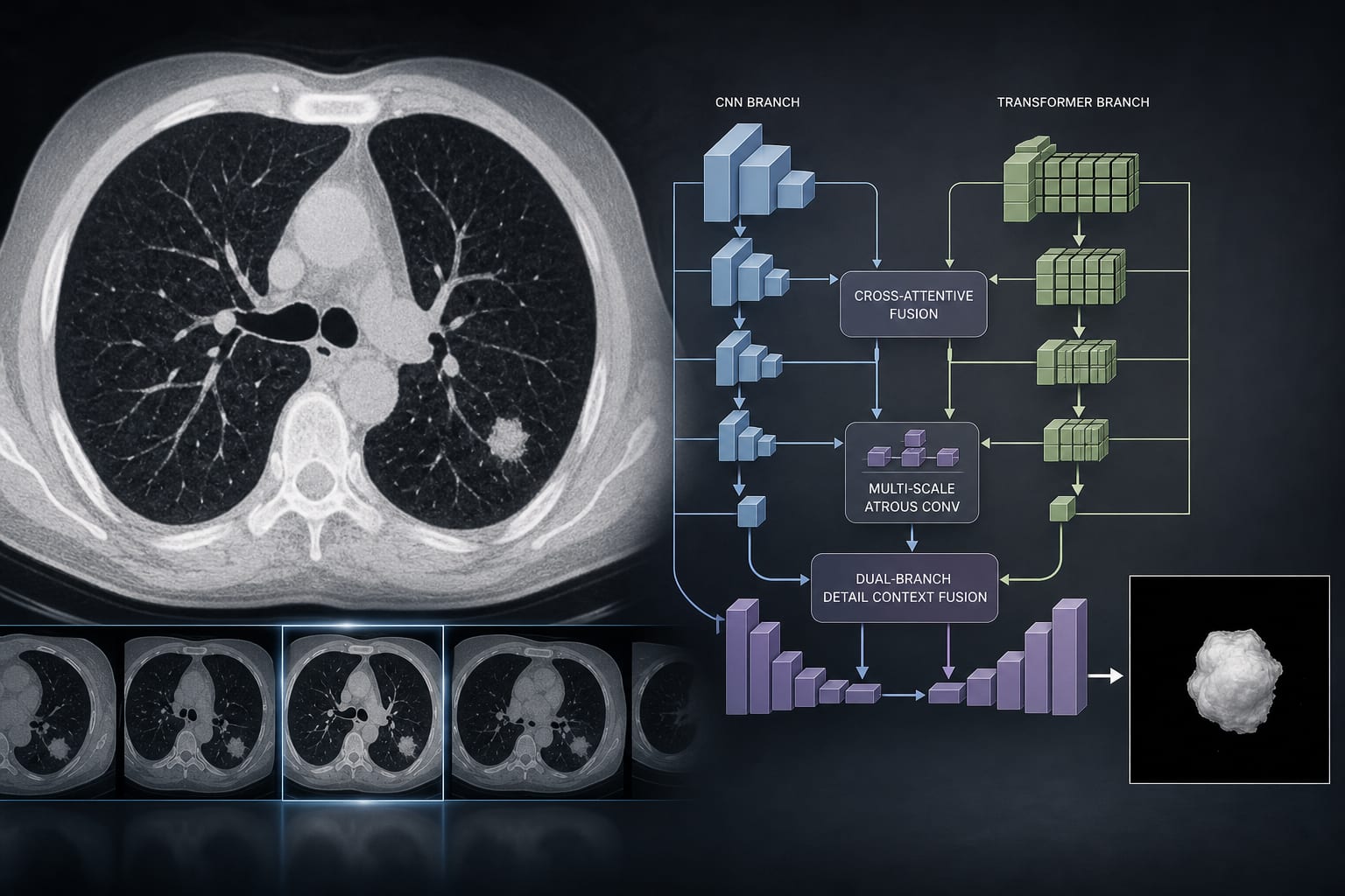
DPCrossU-Net, a dual-branch network combining CNN and Transformer architectures, achieves a Dice score of 85.89% for lung nodule segmentation.
Background
Accurate segmentation of lung nodules in CT images is crucial for early lung cancer detection and diagnosis. The increasing volume of CT data and variability in nodule characteristics pose significant challenges for radiologists.
Data Highlights
No numerical data table provided.
Key Findings
DPCrossU-Net integrates CNN and Vision Transformer representations for enhanced feature extraction.
The model employs a Cross-Attentive Fusion (CAF) module to combine local and global features adaptively.
Multi-scale atrous convolutions improve sensitivity to small nodules.
A dual-branch Detail Context Fusion (DCF) block enhances boundary reconstruction in the decoder.
DPCrossU-Net achieved a Dice score of 85.89% on the LIDC-IDRI dataset, outperforming baseline U-Net models.
Clinical Implications
The DPCrossU-Net model offers a robust solution for lung nodule segmentation, which may facilitate more accurate early lung cancer diagnosis. Its advanced feature extraction capabilities could support the development of more effective CAD systems in clinical settings.
Conclusion
DPCrossU-Net demonstrates significant improvements in lung nodule segmentation accuracy, highlighting the potential of combining CNN and Transformer architectures in medical imaging.