Clinical Report: Uncommon Presentation of a Persistent Proatlantal Intersegmental Artery
Overview
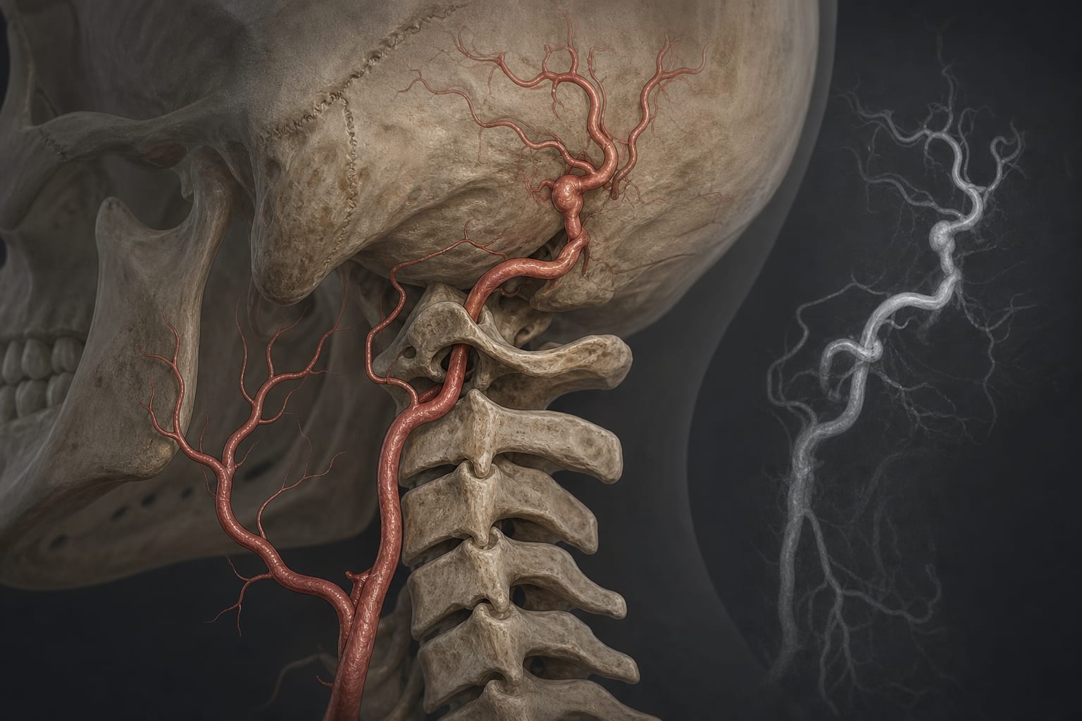
This report details a rare case of a persistent proatlantal intersegmental artery (PPIA) in a 67-year-old male, presenting with recurrent dizziness. The case highlights the importance of recognizing unusual vascular anomalies that can serve as the dominant supply to the posterior circulation, particularly in patients with vertebral artery agenesis.
Background
The persistent proatlantal intersegmental artery (PPIA) is a rare embryonic remnant that connects the carotid and vertebrobasilar systems. Understanding its variations is clinically significant due to potential implications for surgical and interventional procedures. Recognition of PPIA is crucial, especially in patients with vertebral artery agenesis, as it may alter the risk of complications during vascular interventions.
Data Highlights
No numerical data available in the article.
Key Findings
['The patient exhibited a unique vascular anomaly that could not be classified into the classic Lasjaunias types.', 'Imaging revealed a right vertebral artery aneurysm and absence of the left vertebral artery.', 'An anomalous artery from the left external carotid artery supplied the occipital artery and joined the right vertebral artery.', 'This case supports the embryological concept that the occipital artery derives from PIA components.', 'Preoperative identification of such vascular anomalies is essential to avoid complications during surgical interventions.']
Clinical Implications
Clinicians should be aware of the potential for persistent proatlantal intersegmental arteries in patients presenting with dizziness or other neurological symptoms. Preoperative imaging, such as CTA or MRA, is recommended to identify these anomalies and plan accordingly to mitigate risks during surgical procedures.
Conclusion
The case illustrates the importance of recognizing mixed-type PPIAs, which may serve as critical vascular conduits in the posterior circulation. Enhanced awareness and imaging techniques are essential for safe management of such vascular anomalies.