Clinical Report: Multimodal MRI Analysis in Prolonged Disorders of Consciousness
Overview
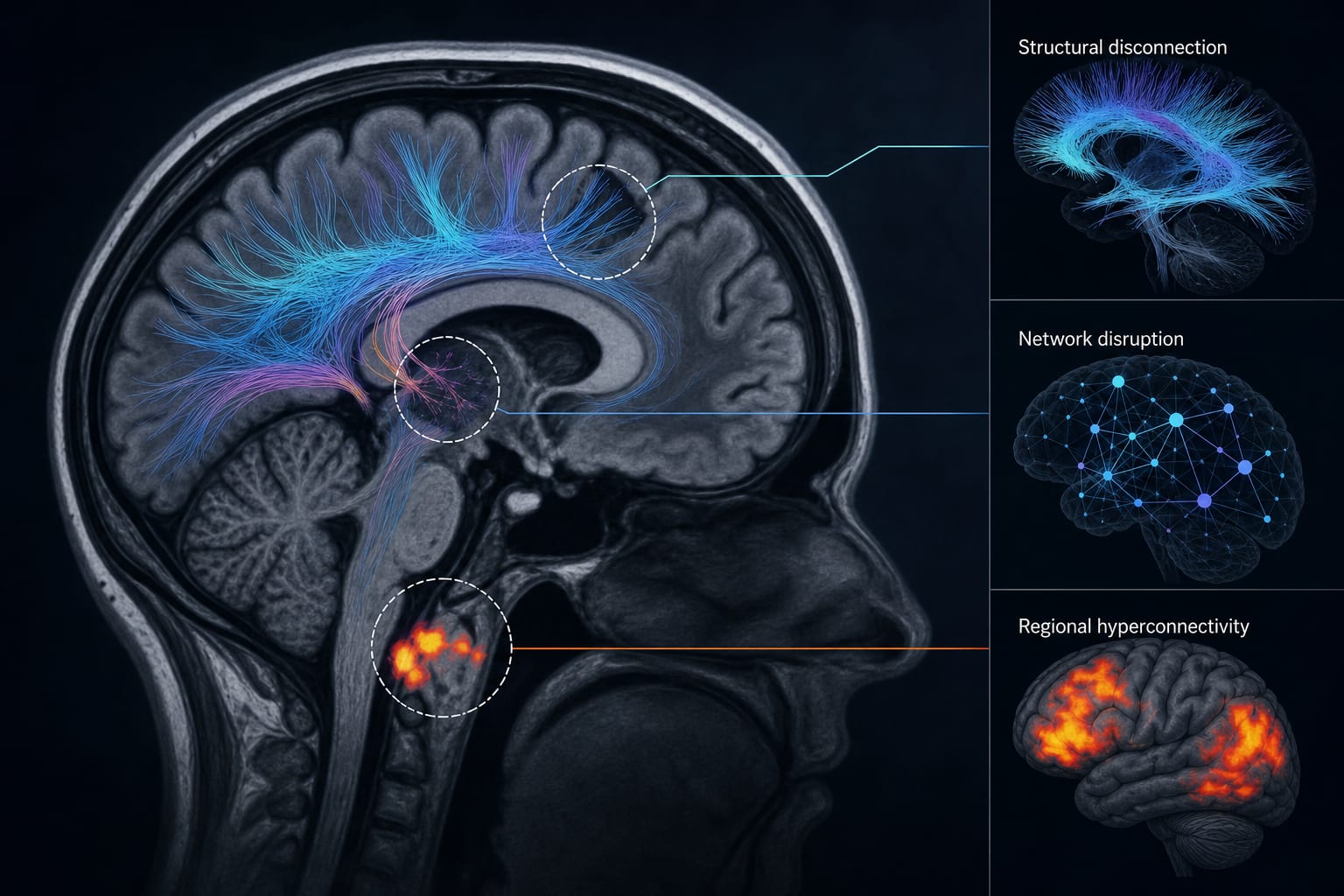
This study identifies three levels of pathological changes in patients with prolonged disorders of consciousness (pDoC) using multimodal MRI techniques. Key findings include structural disconnection, network disruption, and regional hyperconnectivity, which provide insights into the neural mechanisms underlying consciousness impairment.
Background
Prolonged disorders of consciousness (pDoC) present significant diagnostic challenges, as traditional behavioral assessments often fail to detect covert cognitive activity. Accurate diagnosis is crucial for prognosis and treatment planning, as misdiagnosis can lead to inappropriate care. Advances in neuroimaging techniques, particularly multimodal MRI, offer new avenues for understanding and assessing pDoC.
Data Highlights
{'format': 'Ensure the table is properly formatted for clarity.'}
Key Findings
pDoC patients showed reduced fractional anisotropy (FA) in the left anterior corona radiata (ACR-L).
Altered amplitude of low-frequency fluctuations (ALFF), fractional ALFF (fALFF), and regional homogeneity (ReHo) were observed in prefrontal, cerebellar, and limbic regions.
Disrupted functional connectivity (FC) was noted within higher-order cortical networks in pDoC patients.
CRS-R scores positively correlated with ACR-L FA and prefrontal ReHo.
CRS-R scores negatively correlated with hyperactivity in cerebellar and limbic regions.
Clinical Implications
The findings underscore the importance of integrating multimodal neuroimaging in the assessment of pDoC to enhance diagnostic accuracy. Clinicians should consider these neuroimaging biomarkers for better prognostic evaluation and treatment planning.
Conclusion
This study provides a comprehensive framework for understanding the neural mechanisms of consciousness impairment in pDoC, highlighting the potential of multimodal MRI as a diagnostic tool.