Clinical Report: Brain fMRI Responses to Visual Stimulation in Migraine Patients
Overview
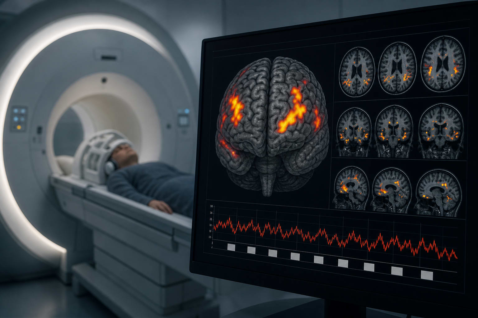
This study investigates brain activation patterns in migraine patients with visual aura (VaM) compared to those without aura (MwoA) and healthy controls (HC). Significant differences in brain activation were observed, particularly in regions associated with visual processing.
Background
Migraine is a prevalent headache disorder that significantly affects quality of life, with visual aura being a common symptom. Misdiagnosis and inadequate treatment often occur due to the complexity of aura symptoms. Understanding the neural correlates of visual aura is important for differential diagnosis.
Data Highlights
Group Comparison
Brain Region
Activation Difference (p-value)
VaM vs HC
Left Precuneus
< 0.005
MwoA vs HC
Right Middle Occipital Gyrus
< 0.005
MwoA vs HC
Left Middle Occipital Gyrus
< 0.05
VaM vs MwoA
Right Angular Gyrus
< 0.05
VaM vs MwoA
Left Cuneus
< 0.0001
VaM vs MwoA
Right Middle Cingulate Gyrus
< 0.05
VaM vs MwoA
Left Precuneus
< 0.05
VaM vs MwoA
Right Middle Occipital Gyrus
< 0.05
Key Findings
21 patients with visual aura migraine (VaM) and 21 with migraine without aura (MwoA) were studied alongside 21 healthy controls.
Significant activation differences were found in brain regions related to visual processing among the three groups.
The VaM group showed stronger activation in the left precuneus compared to healthy controls.
The MwoA group exhibited stronger activation in the right and left middle occipital gyrus compared to healthy controls.
Compared to the MwoA group, the VaM group had stronger activation in the right angular gyrus and left cuneus.
Clinical Implications
The identification of specific brain activation patterns in migraine patients with visual aura may assist in improving diagnostic accuracy. Understanding these neural mechanisms can inform treatment approaches and enhance patient management strategies.
Conclusion
The study highlights distinct brain activation patterns associated with visual aura in migraine patients, warranting further investigation into the underlying mechanisms.
Creatine's ATP-buffering trick isn't just for biceps. In mice, it juices up dendritic cells' energy supply, sharpening T-cell priming and shrinking tumors on daily injections. Human DCs got the memo too, at least in a dish. Early days, but a fun plot twist for gym-bag chemistry.