Subtle changes in the outer retinal and choroidal structures in individuals at elevated risk for advancing to age-related macular degeneration - Report - MDSpire
Subtle changes in the outer retinal and choroidal structures in individuals at elevated risk for advancing to age-related macular degeneration
Overview
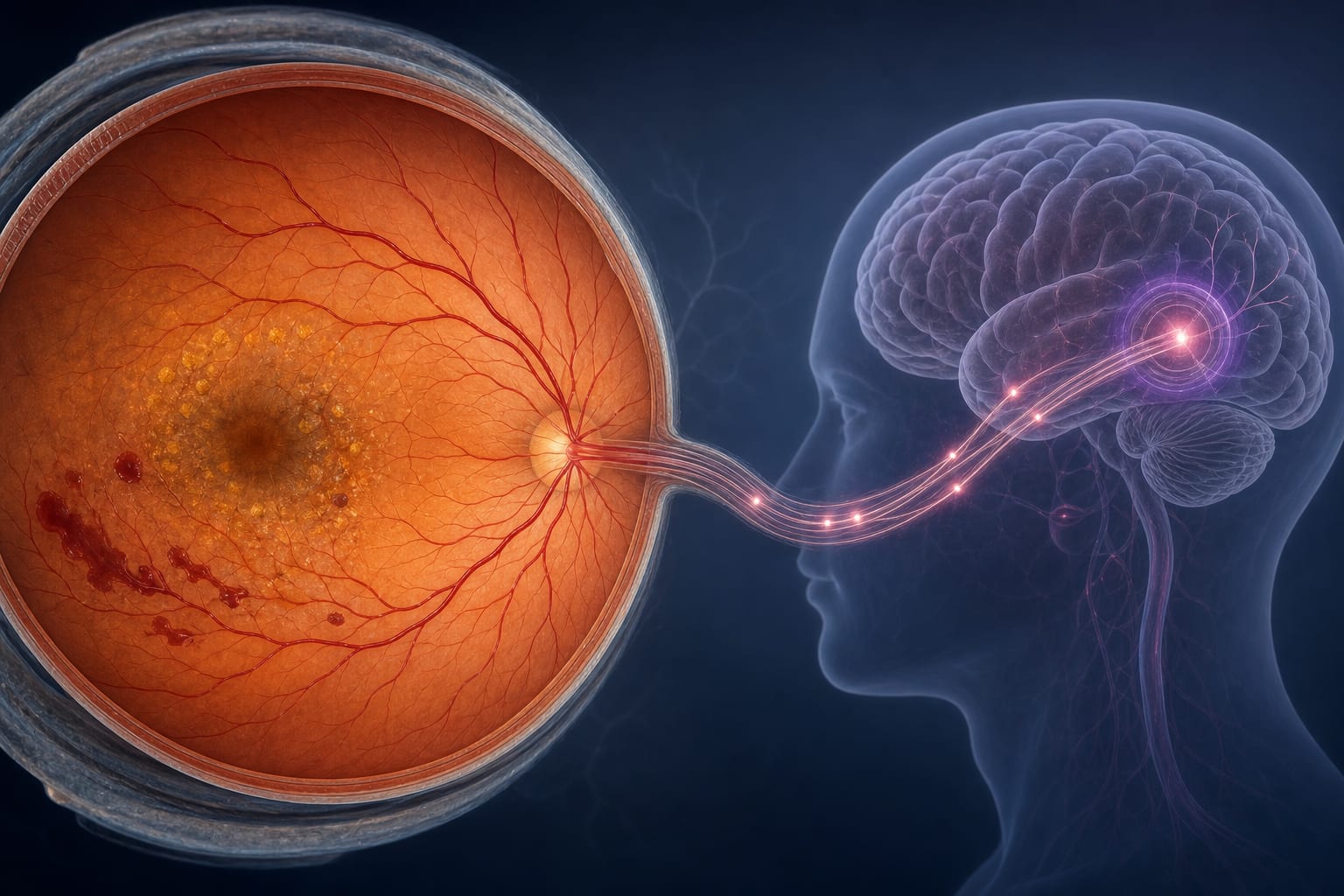
This study investigates microstructural changes in the outer retina and choroidal blood supply in patients at high risk for age-related macular degeneration (AMD). Significant thinning of the outer retina and choroid was observed in patients with dry AMD, indicating potential early biomarkers for disease progression.
Background
Age-related macular degeneration (AMD) is a leading cause of blindness globally, with its prevalence increasing with age. Understanding the early structural changes in the retina and choroid is crucial for timely intervention and management of AMD. The study highlights the importance of advanced imaging techniques like swept-source optical coherence tomography (SS-OCT) in detecting these changes.
Data Highlights
Group
Outer Retinal Thickness
CVI
SCP Density
DCP Density
Controls
Normal
Normal
Normal
Normal
Group 1 (High Risk)
Decreased
Normal
Normal
Normal
Group 2 (Dry AMD)
Significantly Decreased
Significantly Decreased
Significantly Decreased
Significantly Decreased
Key Findings
Significant thinning of outer retinal and choroidal structures in patients with dry AMD compared to controls.
Choroidal vessel index (CVI) was significantly decreased in patients with dry AMD in superior and temporal regions.
Outer retinal thickness was significantly correlated with choroidal thickness in AMD patients.
Microvascular densities of the superficial and deep retinal capillary plexuses were significantly decreased in dry AMD patients.
SS-OCTA is effective in evaluating microstructural and vascular changes in the outer retina.
Clinical Implications
The findings suggest that SS-OCTA can be a valuable tool for early detection of AMD progression by identifying subtle changes in retinal and choroidal structures. Clinicians should consider incorporating this imaging modality into routine assessments for patients at high risk of developing neovascular AMD.
Conclusion
The study underscores the significance of early structural changes in the outer retina and choroid as potential biomarkers for AMD progression. Utilizing advanced imaging techniques may enhance the ability to monitor and manage patients at risk for this debilitating condition.
Roger A. Goldberg, MD, MBA, shared early postapproval experience with Encelto, including an evolving surgical technique, at Clinical Trials at the Summit.