MRI Assessment of Cardiac and Hepatic Iron in Hematological and Liver Diseases
Overview
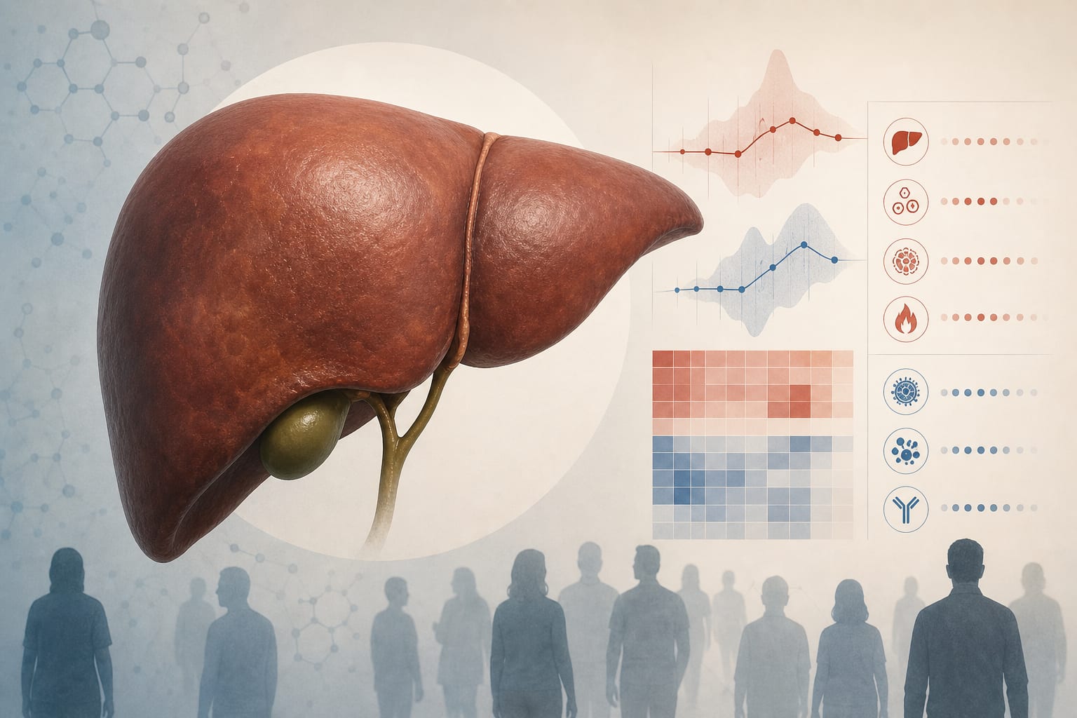
This study evaluated cardiac and liver iron accumulation using MRI in non-chelated patients with hematological malignancies and chronic liver disease. Liver R2 calibrated to MRI-based hepatic iron concentration was used as the reference, revealing variable iron overload patterns and limited cardiac iron deposition except in severe liver iron overload cases.
Background
Iron overload due to transfusions is a major cause of morbidity in hematological diseases, especially thalassemia major where cardiac iron accumulation leads to high mortality. In myelodysplastic syndrome (MDS) and other hematological malignancies, data on myocardial iron deposition are limited, though elevated serum ferritin correlates with worse survival. Chronic liver diseases also show iron accumulation linked to infection risk. Non-invasive MRI techniques measuring T2*, R2, and R2* have emerged as reliable tools for quantifying tissue iron, overcoming limitations of invasive biopsies and nonspecific serum markers.
Data Highlights
Group
Number of Subjects
Mean Age (years)
Imaging Performed
Hematological Patients
75
49.4 ± 13.8
Liver R2 (75), Cardiac T2* (73)
Chronic Liver Disease Patients
12
51.9 ± 11.5
Liver R2, Cardiac T2*
Healthy Volunteers
12
39.4 ± 15.3
Liver R2 (9), Cardiac T2* (12)
Key Findings
Cardiac iron overload was uncommon in non-chelated patients with hematological malignancies and chronic liver disease.
Liver iron concentration measured by MRI R2 correlated well with transfusion history and serum ferritin levels.
Patients with severe liver iron overload showed a higher likelihood of cardiac iron deposition.
Cardiac T2* values demonstrated good reproducibility and were inversely correlated with iron concentration.
Iron chelation therapy alters the relationship between liver and cardiac iron and affects MRI calibration curves.
Non-invasive MRI provides a reliable quantitative assessment of tissue iron, comparable to liver biopsy variability.
Clinical Implications
MRI-based quantification of liver and cardiac iron offers a safe, non-invasive method to monitor iron overload in patients with hematological malignancies and chronic liver disease. This can guide transfusion management and early intervention to prevent cardiac complications. Recognizing that cardiac iron overload is rare except in severe liver iron excess may influence surveillance strategies and chelation decisions.
Conclusion
MRI assessment of iron overload reveals that cardiac iron deposition is infrequent in non-chelated hematological and liver disease patients, except in those with severe hepatic iron accumulation. MRI serves as a valuable tool for comprehensive iron monitoring to optimize patient management.
References
Andersson et al. -- Cardiac T2* MRI Method
Studies on Liver R2 Calibration and MRI Iron Quantification
Research on Iron Overload in MDS and Hematological Malignancies