Clinical Report: Evaluation of Radial Volar Portals in Wrist Arthroscopy
Overview
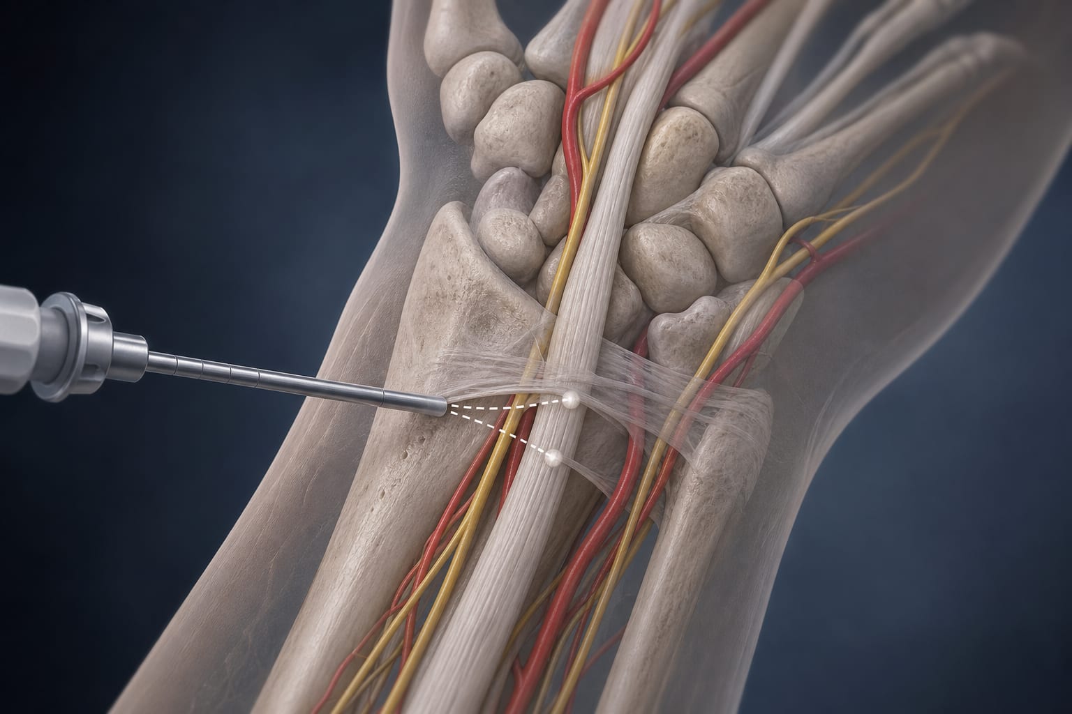
This study evaluates the feasibility and safety of the Kiss-in method for radial volar portals in wrist arthroscopy using 3D CT reconstructions. Significant gender-specific differences in anatomical parameters were identified, particularly concerning the trocar length and the frequency of risky zones near critical structures.
Background
Wrist arthroscopy is essential for diagnosing and treating wrist disorders, yet traditional methods carry risks of neurovascular injury. The Kiss-in method offers a novel approach for portal placement. Understanding the anatomical implications of this technique is crucial for enhancing surgical safety and efficacy.
Data Highlights
Parameter
VRMC Portal
VRRC Portal
dTP
Significant differences between genders
Significant differences between genders
dTF
Significant differences between genders
Not specified
Risky Zone Frequency
Lower
Higher
Key Findings
The Kiss-in method shows theoretical feasibility for establishing radial volar portals.
Statistically significant differences in dTP and dTF were observed between genders.
Female dTP values were consistently below the standard trocar length of 45 mm.
In males, 68.75% of dTP values in the VRRC portal exceeded 45 mm.
The frequency of the 'risky zone' was significantly higher in the VRRC portals.
Clinical Implications
Clinicians should consider anatomical differences when planning wrist arthroscopy procedures.
Conclusion
This imaging study supports the theoretical application of the Kiss-in method for wrist arthroscopy, highlighting important anatomical considerations that may influence surgical outcomes.