Conexiant’s news site is now MDSpire News. Learn more
Advertisement
Imaging Leukocyte Dynamics in Retinal Disease
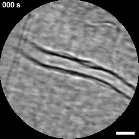
The movement of immune cells through the retinal vasculature has long been inferred from laboratory models. Now, advances in imaging are allowing clinicians to observe these processes directly. At the 2026 meeting of the Association for Research in Vision and Ophthalmology (ARVO) in Denver, Richard B. Rosen, MD, ScD(hon), FACS, FASRS, FARVO, described early clinical work using adaptive optics scanning laser ophthalmoscopy (AOSLO) to visualize leukocyte trafficking in vivo.
Clinical Report: Imaging Leukocyte Dynamics in Retinal Disease
Overview
This report discusses the use of adaptive optics scanning laser ophthalmoscopy (AOSLO) to visualize leukocyte trafficking in retinal diseases. Findings indicate that leukocyte behavior varies across different retinal conditions, providing insights into immune responses and potential treatment effects.
Background
Understanding leukocyte dynamics in the retina is crucial for elucidating the immune mechanisms underlying various retinal diseases. Advances in imaging technology, such as AOSLO, allow for direct observation of these processes, which were previously inferred from laboratory models. This capability may enhance our understanding of disease progression and treatment responses.
Data Highlights
Condition
Participants
Leukocyte Behavior Observed
Healthy
16
Normal crawling patterns
Diabetic Retinopathy
5
Stalling and adhesion near nonperfusion areas
Sickle Cell Disease
2
Adhesion and occlusive behavior
Uveitis
5
Increased extravascular activity
Key Findings
AOSLO enables noninvasive visualization of leukocyte dynamics in vivo.
Leukocyte activity is more frequently observed in venules compared to arterioles, with a 10:1 difference.
Cell behavior is consistent across vessel sizes, with limited variation in measured velocities.
Distinct leukocyte behaviors were noted in various retinal diseases, including stalling in diabetic retinopathy and increased activity in uveitis.
Changes in leukocyte dynamics may occur following anti-VEGF therapy.
Clinical Implications
The ability to visualize leukocyte dynamics may enhance our understanding of retinal disease mechanisms and inform therapeutic strategies. Clinicians may consider these dynamics when evaluating treatment responses and disease progression.
Conclusion
In vivo imaging of leukocyte trafficking offers a promising avenue for advancing our understanding of retinal diseases and their treatment. Further research is needed to clarify the implications of these findings for clinical practice.