Development and validation of a nomogram based on tumor margin irregularity and alpha-fetoprotein for predicting microvascular invasion in hepatocellular carcinoma - Report - MDSpire
Advertisement
Development and validation of a nomogram based on tumor margin irregularity and alpha-fetoprotein for predicting microvascular invasion in hepatocellular carcinoma
Clinical Report: Nomogram for Predicting Microvascular Invasion in HCC
Overview
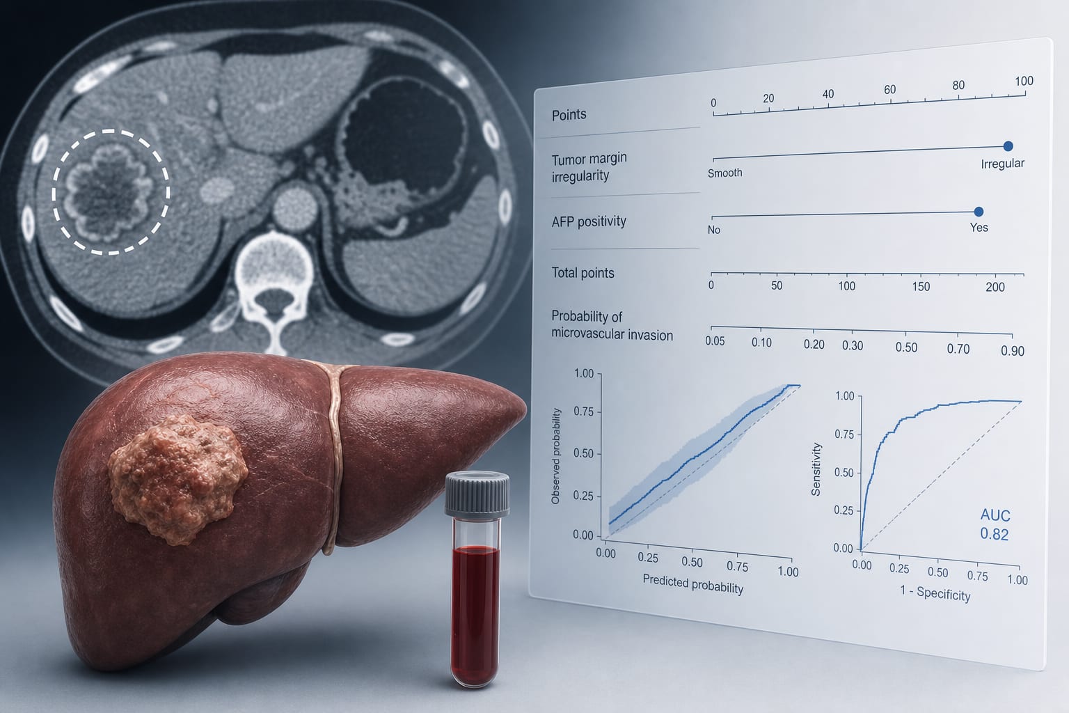
A preoperative nomogram utilizing tumor margin irregularity and alpha-fetoprotein (AFP) levels was developed and validated for predicting microvascular invasion (MVI) in hepatocellular carcinoma (HCC). The model demonstrated discrimination in both training and validation cohorts.
Background
Hepatocellular carcinoma (HCC) is a leading cause of cancer-related mortality globally. Microvascular invasion (MVI) is a significant histopathological marker associated with aggressive HCC behavior and poor prognosis. Identifying patients at risk for MVI preoperatively can enhance surgical planning and postoperative management.
Data Highlights
Cohort
MVI Present
Total Patients
Training
187
487
Validation
95
256
Key Findings
MVI was present in 187 of 487 patients in the training cohort.
MVI was present in 95 of 256 patients in the validation cohort.
Irregular tumor margins and AFP positivity were identified as independent predictors of MVI.
The final nomogram achieved AUCs of 0.740 and 0.781 in the training and validation cohorts, respectively, as reported in the study.
Calibration of the model was acceptable.
Clinical Implications
Further prospective validation is necessary to confirm the findings of this study.
Conclusion
The developed nomogram based on tumor margin irregularity and AFP levels provides a tool for predicting MVI in HCC.