Clinical Report: Utilizing Immersive 3D Visualization in Congenital Heart Surgeries
Background
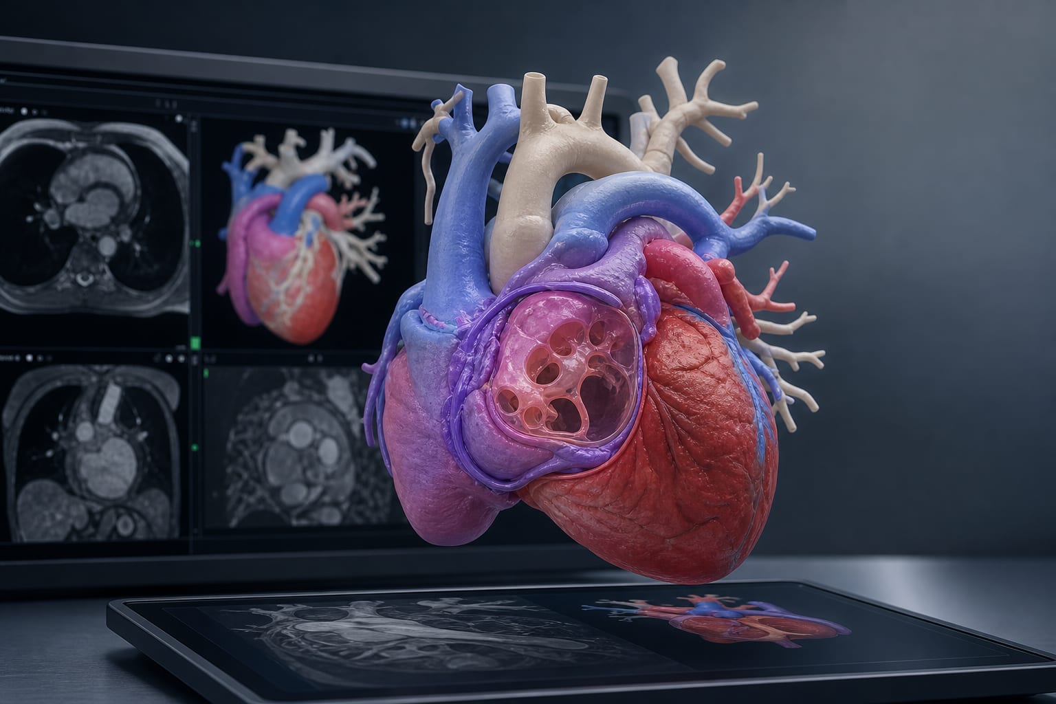
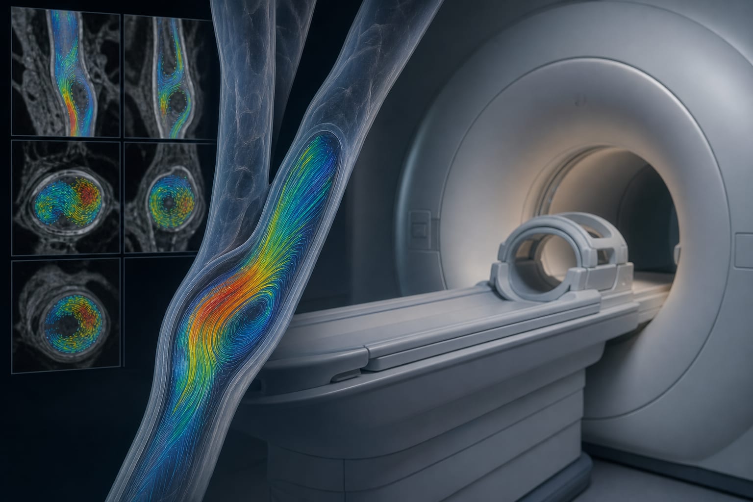
Complex congenital cardiac surgeries present significant challenges due to intricate spatial relationships within the heart. Traditional two-dimensional imaging methods may not suffice for planning these complex cases. The adoption of advanced three-dimensional visualization techniques could enhance preoperative planning and team communication.
Data Highlights
No numerical data or trial results were provided in the source material.
Key Findings
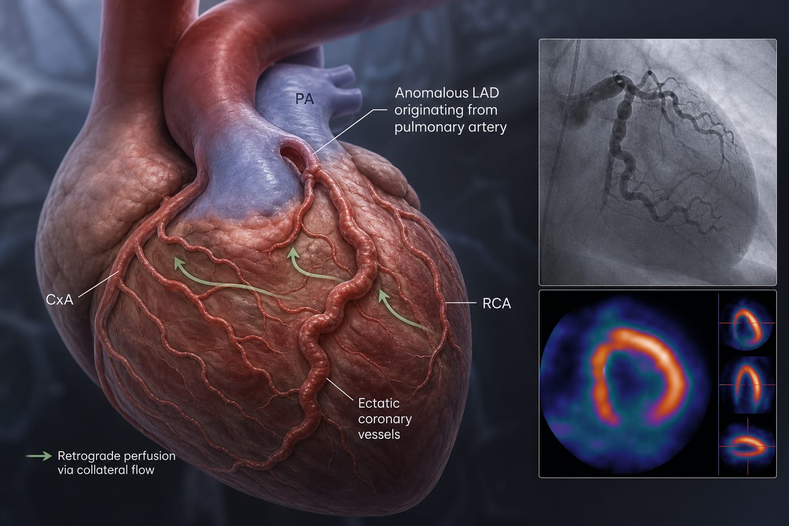
Three-dimensional models improve comprehension of complex congenital anatomy.
Surgeons report increased preoperative confidence and reduced intraoperative anatomical reorientation after reviewing 3D models.
Educational outcomes show that trainees using VR or 3D models perform better in anatomical assessments compared to traditional 2D methods.
Current evidence linking 3D visualization to reduced operative time and complications is limited and requires further study.
Specific scenarios for 3D visualization use include complex ventricular-arterial anomalies and reoperative sternotomies.
Clinical Implications
Clinicians should consider utilizing these technologies in cases where standard imaging leaves uncertainty about critical anatomical relationships.
Conclusion
The integration of immersive 3D visualization in congenital heart surgery planning represents a potential advancement.