Primary central nervous system lymphoma and tumefactive demyelinating lesions in multiple sclerosis: a retrospective observational cohort study - Report - MDSpire
Advertisement
Primary central nervous system lymphoma and tumefactive demyelinating lesions in multiple sclerosis: a retrospective observational cohort study
Differentiating Primary Central Nervous System Lymphoma from Tumefactive Demyelinating Lesions
Overview
This study compares clinical, neuroimaging, and pathological features between primary central nervous system lymphoma (PCNSL) and tumefactive demyelinating lesions (TDLs). Key findings indicate significant differences in age of onset, MRI characteristics, and biomarker levels.
Background
Differentiating PCNSL from TDLs is crucial due to their similar clinical presentations and imaging features, which can lead to misdiagnosis. Accurate diagnosis is essential as treatment approaches differ significantly between these conditions.
Data Highlights
Feature
PCNSL
TDLs
Mean Age
59 ± 10 years
36 ± 8 years
CD20 Staining
Strongly positive
Lower levels
CD3 Staining
Negative
Stronger levels
CSF IL-6 Levels
Higher
Lower
CSF IL-10 Levels
Higher
Lower
Key Findings
PCNSL patients had a significantly older mean age of onset compared to TDL patients (59 vs. 36 years).
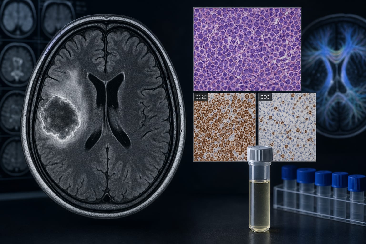
PCNSL lesions exhibited U-shaped hyperintensity on MRI, while TDL lesions showed slightly diffuse hyperintensity.
Pathological analysis revealed strong CD20 positivity in PCNSL and lower CD20 levels in TDLs.
CSF levels of IL-6 and IL-10 were significantly higher in PCNSL compared to TDLs.
PCNSL lesions showed marked perifocal edema on FLAIR sequences, contrasting with isointense TDL lesions.
Clinical Implications
Clinicians should consider age, MRI characteristics, and specific biomarker levels when differentiating between PCNSL and TDLs. Accurate diagnosis is vital to avoid inappropriate treatments that could adversely affect patient outcomes.
Conclusion
A comprehensive evaluation of clinical, neuroimaging, and pathological features, along with biomarker analysis, is essential for the differential diagnosis between PCNSL and TDLs.