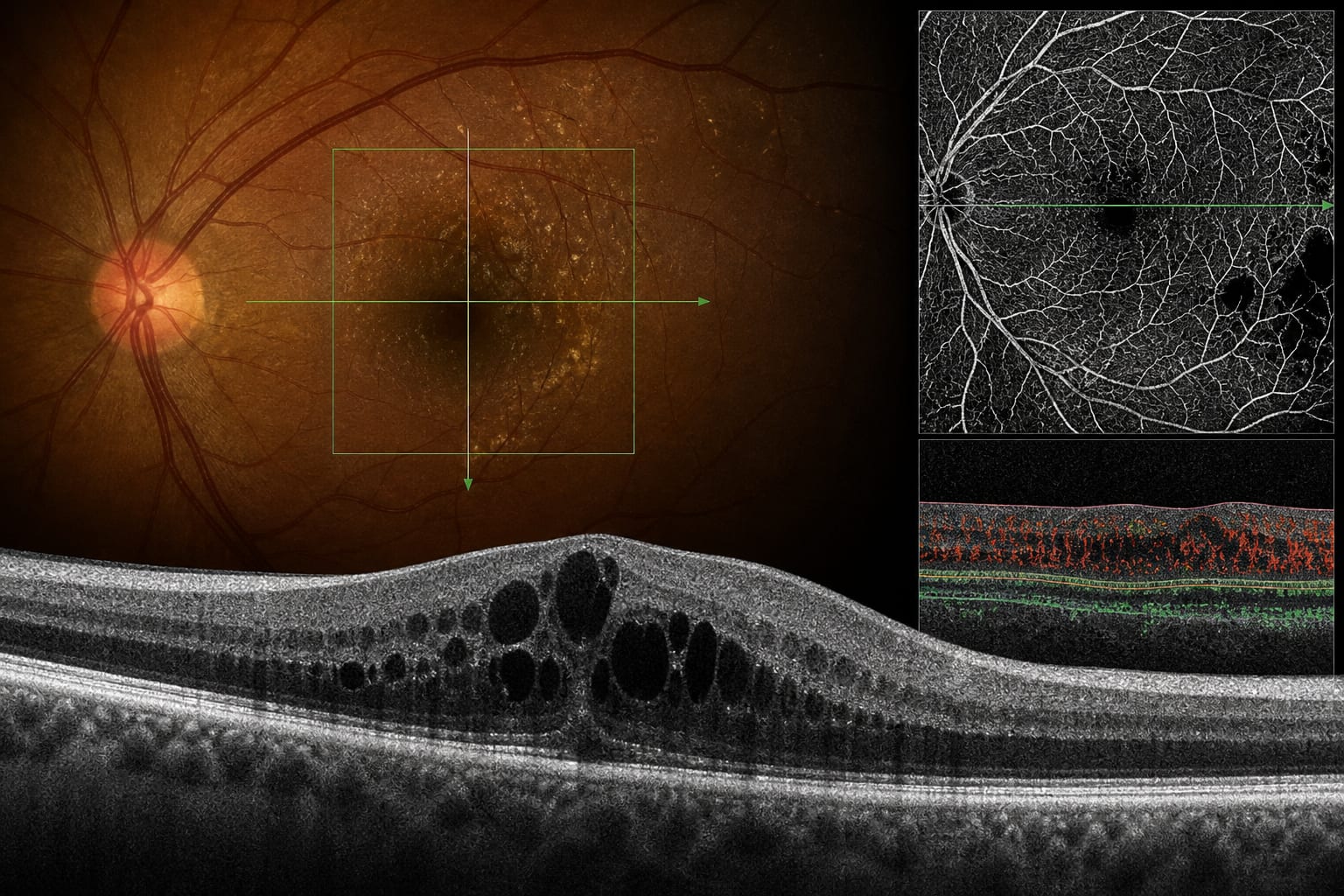
Imaging markers for diabetic macular oedema: Forecasting visual outcomes beyond retinal thickness
-
By
-
Enrico Borrelli
-
October 18, 2026
Clinical Report: Imaging markers for diabetic macular oedema
Overview
In a post hoc analysis of 60 KINGFISHER trial eyes, baseline central subfield thickness and an OCTA composite of superficial perfusion density and vessel length density were independently associated with week-52 visual acuity; baseline hyperreflective foci volume was associated with a smaller visual-acuity gain. The findings support investigation of structural and vascular imaging markers beyond retinal thickness, while the study’s sample size, optional OCTA acquisition, device restriction, and model limitations constrain interpretation.
Background
Anti-VEGF therapy has altered the natural history of diabetic macular oedema (DMO), but individual visual outcomes remain difficult to predict. Central subfield thickness explains only a limited proportion of visual-acuity variation, and anatomical and functional outcomes may not correspond closely. Structural OCT studies have evaluated markers including disorganisation of the retinal inner layers, external limiting membrane and ellipsoid zone integrity, and intraretinal hyperreflective foci. OCT angiography (OCTA) also enables quantitative assessment of retinal capillary networks and diabetic macular ischaemia across separate capillary plexuses.
Data Highlights
| Measure or finding | Reported result |
|---|---|
| Study population | 60 eyes from the KINGFISHER trial with available baseline spectral-domain OCT and OCTA measurements |
| Mean visual acuity | 62.9 letters at baseline; 75.4 letters at week 52 |
| Baseline visual acuity predictor | Greater baseline central subfield thickness was the only independent predictor reported |
| Week-52 visual acuity associations | Baseline central subfield thickness and a composite of superficial perfusion density and vessel length density were independently associated |
| Visual-acuity gain association | Higher baseline hyperreflective foci volume was independently associated with a smaller gain |
| Model variance explained | 19%–32%; models were not adjusted for multiple comparisons |
| Systematic review | 27 studies evaluated 75 candidate biomarkers; five were associated with reduced visual acuity at two or more time points, with moderate certainty of evidence |
Key Findings
- In the KINGFISHER post hoc analysis, mean visual acuity increased from 62.9 letters at baseline to 75.4 letters at week 52.
- Greater baseline central subfield thickness was the only independent predictor of baseline visual acuity reported in the analysis.
- Baseline central subfield thickness and a composite index of superficial perfusion density and vessel length density were independently associated with week-52 visual acuity.
- Higher baseline hyperreflective foci volume was independently associated with a smaller visual-acuity gain.
- A systematic review and meta-analysis of 27 studies and 75 candidate biomarkers found five associated with reduced visual acuity at two or more time points, with moderate certainty of evidence; long-term investigations identified DRIL and outer retinal alterations as features closely associated with vision after years of anti-VEGF therapy.
- The source reports important limitations: OCTA was not required by the parent protocol, analysis was restricted to Heidelberg Spectralis scans, models explained 19%–32% of variance, and multiple comparisons were not adjusted for.
Clinical Implications
The reported associations identify OCT and OCTA measures for further prognostic evaluation, but the source does not establish transferable clinical thresholds or individual-level prediction. OCTA measurements depend on device, scan protocol, segmentation and slab definitions, artefact removal, and binarisation methods, so measurements and thresholds may not be interchangeable across centres.
Conclusion
The analysis adds OCTA vascular measures and hyperreflective foci volume to the imaging features associated with visual outcomes in DMO. Larger, prespecified imaging studies and consistent measurement methods are needed to validate these exploratory associations.
Related Resources & Content
- Alhelaly et al., KINGFISHER post hoc analysis, year not stated -- Imaging markers for diabetic macular oedema: Forecasting visual outcomes beyond retinal thickness
- Systematic review and meta-analysis, year not stated -- Baseline OCT Biomarkers Associated with Visual Acuity in Diabetic Macular Edema
- Retinal Physician, 2020 -- Disorganization of the Retinal Inner Layers as a Prognostic Biomarker
- American Diabetes Association, 2026 -- Retinopathy, Neuropathy, and Foot Care: Standards of Care in Diabetes—2026
- American Academy of Ophthalmology, 2025 -- Diabetic Retinopathy Preferred Practice Pattern
- British Journal of Ophthalmology — AI-Driven Fluid Measurement Forecasts Clinical Results in Eyes with Diabetic Macular Oedema Undergoing Intravitreal Dexamethasone Implant Treatment: Insights from the DIADEMA Study
- Frontiers in Medicine — Phenotype-driven management of diabetic macular edema: multimodal imaging biomarkers and individualized therapy
- Retinal Physician — Novel Methods and Diagnostic Tools in Diabetic Retinopathy Novel Methods and Diagnostic Tools in Diabetic Retinopathy Recommendations
- Baseline OCT Biomarkers Associated with Visual Acuity in Diabetic Macular Edema: A Systematic Review and Meta-analysis
- Disorganization of the Retinal Inner Layers as a Prognostic Biomarker
- Retinopathy, Neuropathy, and Foot Care: Standards of Care in Diabetes—2026
- Diabetic Retinopathy Preferred Practice Pattern
- 12. Retinopathy, Neuropathy, and Foot Care: Standards of Care in Diabetes—2026 | Diabetes Care | American Diabetes Association
- Diabetic Retinopathy Preferred Practice Pattern® - Ophthalmology
- Aflibercept Monotherapy or Bevacizumab First for Diabetic Macular Edema | New England Journal of Medicine
- INFOGRAPHIC: 2-year efficacy, durability and safety of intravitreal faricimab with treat-and-extend dosing up to 16 weeks in diabetic macular oedema (pooled results from YOSEMITE and RHINE) | Eye
- Intravitreal Aflibercept 8 mg for Diabetic Macular Edema - Ophthalmology
- Baseline OCT Biomarkers Associated with Visual Acuity in Diabetic Macular Edema: A Systematic Review and Meta-analysis - PubMed
- OCT outcomes as biomarkers for disease status, visual function, and prognosis in diabetic macular edema
- Assessment of Baseline Ultrawidefield Fluorescein Angiographic Quantitative Leakage Parameters with Ultrawidefield Fundus Features and Clinical Parameters in Diabetic Retinopathy in Protocol AA - PubMed
Based on findings from:
Imaging biomarkers in diabetic macular oedema: Predicting vision beyond retinal thickness
Enrico Borrelli. Eye, 2026.
https://www.nature.com/articles/s41433-026-04986-0
This content is an AI-generated, fully rewritten summary based on a published scholarly article. It does not reproduce the original text and is not a substitute for the original publication. Readers are encouraged to consult the source for full context, data, and methodology.