Deep learning reconstruction improves detection of focal liver lesions in hepatobiliary phase compared to conventional EOB-MRI - Report - MDSpire

Deep learning reconstruction improves detection of focal liver lesions in hepatobiliary phase compared to conventional EOB-MRI

  • By

  • Xuewen Peng

  • Qichao Cheng

  • Runzhe Tian

  • Linlin Lang

  • Cheng Li

  • Ruixin Tao

  • Tianyong Xu

  • Dmytro Pylypenko

  • Liping Zuo

  • Dexin Yu

  • Weiwei Lv

  • May 1, 2026

  • 0 min

Share

Clinical Report: Enhanced Detection of Focal Liver Lesions Using DL Recon

Overview

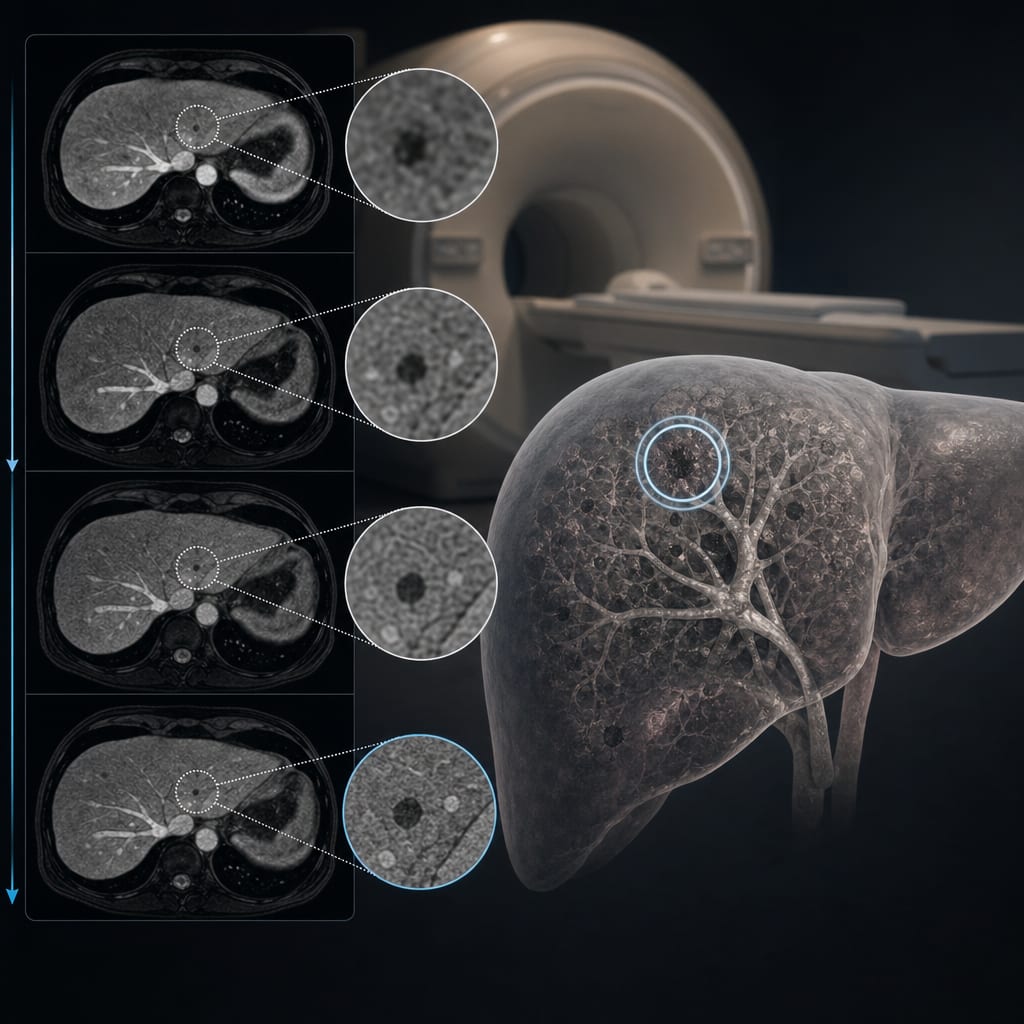
This study evaluates the efficacy of Deep Learning Reconstruction (DL Recon) in enhancing the detection of small focal liver lesions (FLLs) during the hepatobiliary phase of EOB-MRI. Results indicate that DL Recon significantly improves the identification of small low-signal lesions compared to standard non-DL techniques.

Background

The detection of small focal liver lesions (FLLs) is crucial for early diagnosis and treatment of liver diseases, including hepatocellular carcinoma. Conventional MRI techniques often struggle with small lesions due to limitations in signal-to-noise ratio and prolonged scan times. The advent of Deep Learning Reconstruction (DL Recon) technology presents a promising solution to enhance imaging quality and lesion detectability.

Data Highlights

{'table': {'rows': [{'lesion_size': '5-10 mm', 'standard_dl': 'More lesions detected', 'standard_non_dl': 'Fewer lesions detected', 'p_value': '0.030'}, {'lesion_size': '<5 mm', 'standard_dl': 'More lesions detected', 'standard_non_dl': 'Fewer lesions detected', 'p_value': '<= 0.032'}]}}

Key Findings

  • DL Recon shows significant improvement in detecting small low-signal lesions compared to standard non-DL methods.
  • High-signal lesions did not show significant differences among the imaging groups.
  • Intraclass correlation coefficient (ICC) among all groups was >0.90, indicating excellent consistency.
  • For lesions >10 mm, no notable differences were found between DL and non-DL groups.
  • DL methods significantly increased the detection rate of lesions <10 mm.

Clinical Implications

The findings suggest that incorporating DL Recon in EOB-MRI can enhance the detection of small liver lesions, potentially leading to earlier diagnosis and improved patient outcomes. Radiologists should consider utilizing DL techniques to optimize imaging protocols for liver assessments.

Conclusion

The integration of Deep Learning Reconstruction in EOB-MRI significantly enhances the detection of small focal liver lesions, underscoring its clinical relevance in liver imaging.
References

  1. European Radiology, 2023 -- Association of Histological, Imaging, and AI Characteristics in Patients with NAFLD Using Gd-EOB-DTPA-Enhanced MRI: A Proof-of-Concept Investigation
  2. European Radiology, 2024 -- Improving Liver MRI with Gadoxetic Acid: A Combined Method Utilizing Deep Learning CAIPIRINHA-VIBE and Enhanced Fat Suppression Techniques
  3. European Radiology, 2025 -- Utilizing Deep Learning Techniques for Identifying Liver Lesions in Standard and Low-Dose Abdominal CT Scans
  4. The American College of Radiology -- Clinical Practice Guidelines for MR of the Liver
  5. European Radiology — Models for Distinguishing Benign and Malignant Liver Lesions Using Multiparametric Dual-Energy Non-Contrast CT
  6. ACR Practice Parameter for MR of the Liver
  7. Diagnostic value of Gd-EOB-DTPA-enhanced MRI versus contrast-enhanced CT for detecting liver metastasis in colorectal cancer: a systematic review and meta-analysis | BMC Gastroenterology | Springer Nature Link
  8. AI-augmented reconstruction provides improved image quality and enables shorter breath-holds in contrast-enhanced liver MRI

Original Source(s)

Related Content