Clinical Report: Cellular and Stromal Changes Contributing to Heart Damage from Radiation
Overview
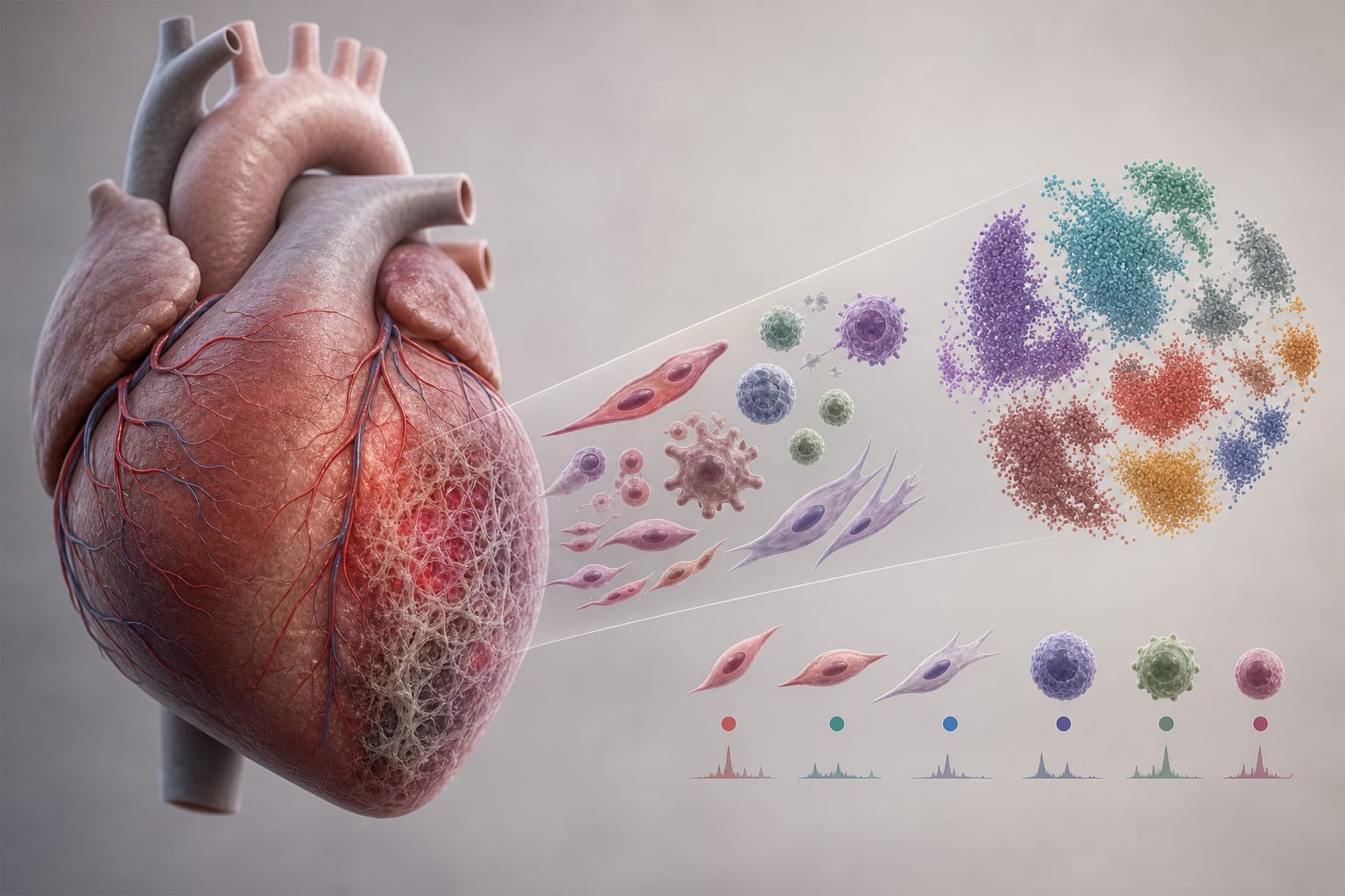
This study utilizes single-cell RNA sequencing to explore the cellular and molecular mechanisms of radiation-induced heart injury (RIHI). Key findings reveal significant alterations in endothelial cells and fibroblasts.
Background
Radiation-induced heart injury (RIHI) is a significant late complication of thoracic radiotherapy, affecting cardiovascular health in cancer survivors. The mechanisms underlying RIHI are complex and involve interactions among various cell types, leading to myocardial damage.
Data Highlights
Single-cell RNA sequencing was performed on rat hearts and matched peripheral blood mononuclear cells (PBMCs) 12 weeks post-irradiation, profiling 38,941 cardiac cells and 41,097 PBMCs.
Key Findings
Endothelial cells (ECs) exhibited subtype shifts and marked MHC-II upregulation following irradiation.
Fibroblasts showed iron accumulation and pro-inflammatory activation, along with antigen-presenting properties.
Myeloid activation was observed, with macrophage and IL-1β⁺ neutrophil programs being prominent.
T and NK cells polarized toward cytotoxic states, indicating immune system involvement in RIHI.
B cells enhanced antigen presentation, contributing to the chronic inflammation observed in RIHI.
Clinical Implications
The findings suggest that targeting non-hematopoietic antigen presentation may be a viable therapeutic strategy in managing RIHI. This approach could be particularly effective when combined with immune checkpoint inhibitors during thoracic radiotherapy.
Conclusion
The study delineates a stromal-immune cascade in RIHI, highlighting the role of endothelial and fibroblast reprogramming.