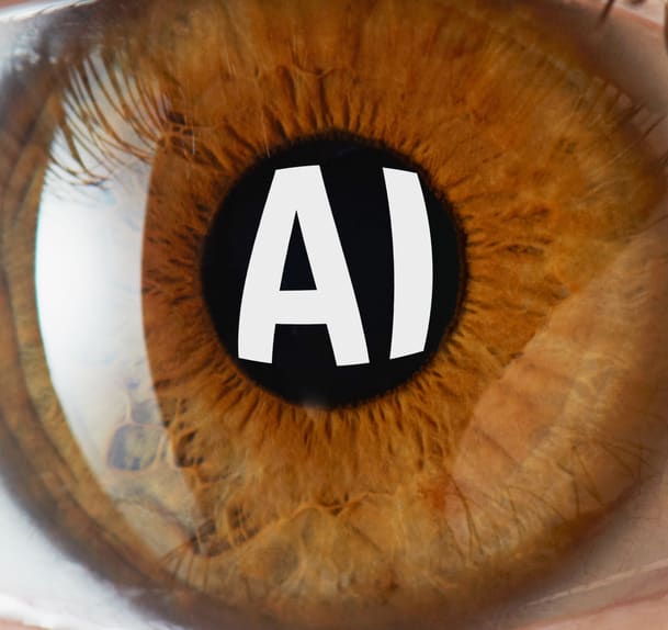
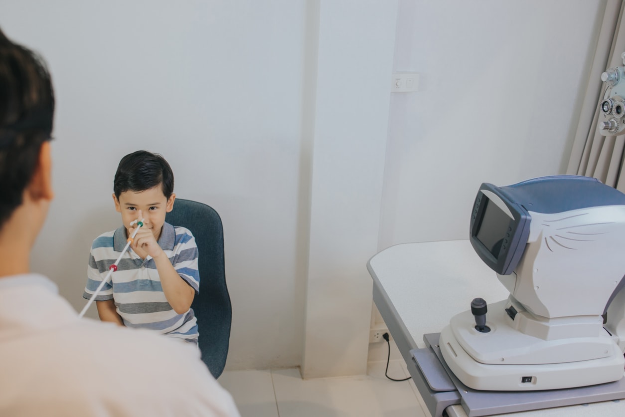
Clinical Report: Retinal Imaging AI May Flag Brain Health Risk
Overview
AI-driven analysis of retinal images shows potential for early risk stratification for dementia and stroke, although current evidence does not confirm improvements in clinical decisions or patient outcomes.
Background
The ability to assess brain health through retinal imaging is significant because the retina shares characteristics with the central nervous system and can be visualized noninvasively.
Data Highlights
No numerical data provided in the source material.
Key Findings
AI-driven 'oculomics' may assess brain health via retinal imaging.
Prior studies suggest AI models can detect Alzheimer dementia and early cognitive impairment from retinal images.
AI models have identified silent brain infarctions from retinal photographs, improving stroke prediction.
Most oculomics models are in early clinical evaluation and judged by technical performance metrics.
Clear definitions of intended use and clinical settings are necessary for broader deployment of oculomics.
Clinical Implications
Clinicians should remain informed about the evolving role of oculomics in risk assessment and management.
Conclusion
Further research is needed to establish the clinical utility and effectiveness of AI-driven retinal imaging.
Deseyne delivers smooth, continuous focus across near, intermediate, and distance vision, which is enabled by Cataltheia’s patented hyper-refractive central zone, engineered to precisely redirect light in a controlled manner.