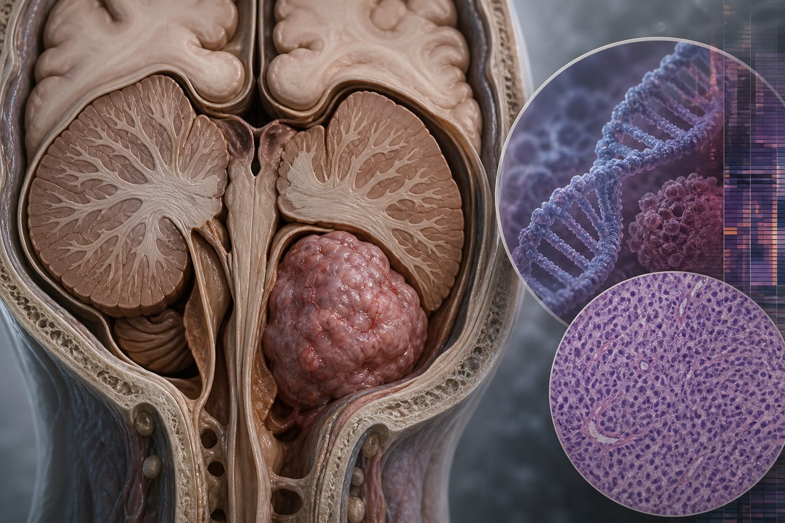
Posterior fossa ependymoma: a comprehensive review of molecular classification, management guidelines, and clinical outcomes (Part I of ependymomas across compartments) - Report - MDSpire
Conexiant’s news site is now MDSpire News. Learn more
Advertisement
Ependymomas in the Posterior Fossa: An In-Depth Analysis of Molecular Classification, Treatment Protocols, and Clinical Outcomes (Part I on Ependymomas in Various Locations)
Clinical Report: Ependymomas in the Posterior Fossa: An In-Depth Analysis
Background
Ependymomas are rare central nervous system tumors, particularly in children, where they account for about 5% of pediatric CNS tumors. The posterior fossa is a common site for these tumors, and nearly 45% of affected children experience recurrence, complicating treatment.
Data Highlights
No numerical data available in the source material.
Key Findings
Ependymomas are classified into two main molecular subgroups: PFA and PFB, which have distinct biological and clinical features.
PFA tumors are associated with poorer clinical outcomes and are more common in younger children, with a median age of 5 years.
PFB tumors typically present in adolescents and adults and exhibit a less aggressive clinical course.
Epigenetic changes, such as H3K27me3 loss, are significant in PFA tumors and serve as important diagnostic markers.
1q gain and/or 6q loss in PFA tumors indicate a very high-risk subset linked to recurrence and poor survival.
MRI is the primary imaging modality for assessing PFE, with distinct imaging phenotypes observed between PFA and PFB tumors.
Clinical Implications
Molecular classification of ependymomas is essential for risk stratification and treatment planning. Understanding the differences between PFA and PFB can guide clinicians in managing these tumors more effectively.
Conclusion
The integration of molecular profiling into the classification of posterior fossa ependymomas enhances prognostic accuracy and informs treatment strategies, ultimately aiming to improve clinical outcomes.