Clinical Report: Utilizing Machine Learning and Resting-State fMRI in PSD
Overview
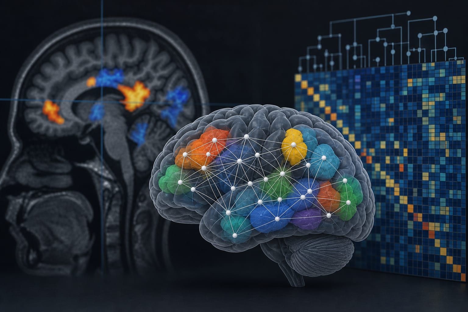
This study identifies resting-state functional differences in patients with post-stroke depression (PSD) compared to healthy controls, utilizing machine learning to analyze imaging features.
Background
Post-stroke depression (PSD) affects a significant proportion of stroke survivors, impacting recovery and quality of life. Understanding the neurobiological underpinnings of PSD through advanced imaging techniques like resting-state functional MRI (rs-fMRI) can provide insights into its characteristics.
Data Highlights
Feature Type
Count
ReHo
7
ALFF
8
DC
6
FC
8
Key Findings
Patients with PSD exhibited resting-state functional differences in multiple brain regions.
A total of 29 candidate features showed significant differences between PSD patients and healthy controls.
LASSO regression identified 10 core features with a cross-validated AUC of 0.878.
The Extra Trees model achieved the highest independent test-set performance with an AUC of 0.889.
SHAP analysis revealed key features influencing the model, including DC in the left anterior cingulate and ReHo in the left thalamus.
Clinical Implications
The identification of specific resting-state functional differences in PSD patients may guide future diagnostic and therapeutic strategies. Clinicians should consider these neurobiological variations when assessing and managing depression in stroke survivors.
Conclusion
The study provides insights into the functional brain differences associated with PSD. Further validation in larger cohorts is necessary.