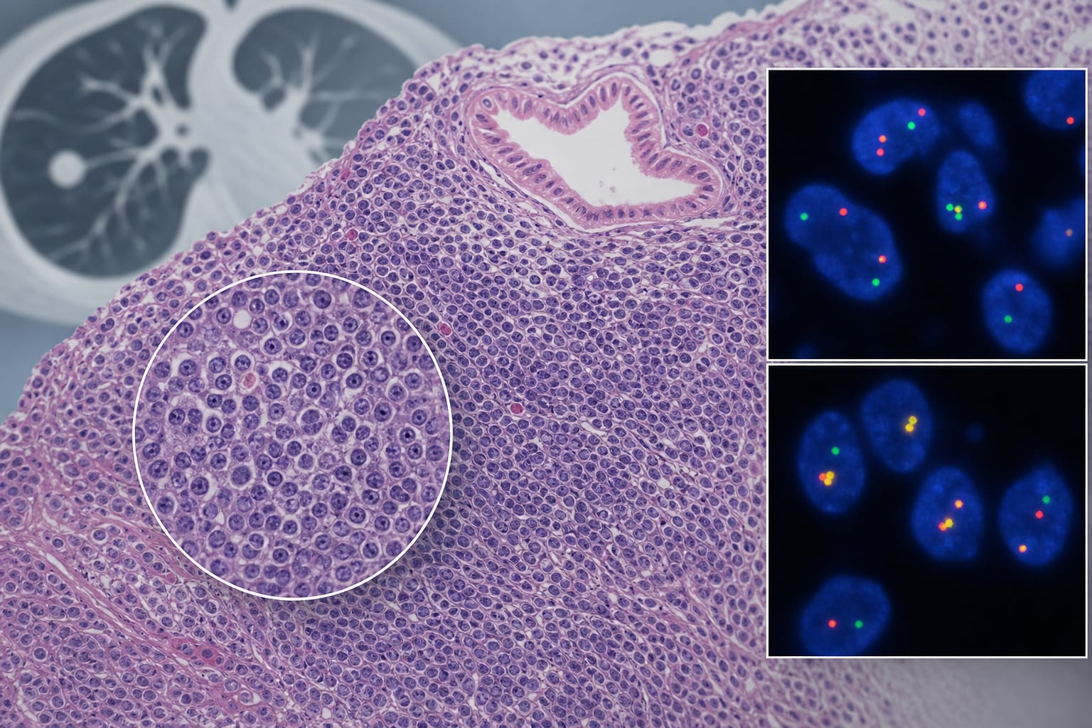
This case study presents a 49-year-old man diagnosed with pulmonary MALT lymphoma featuring a BIRC3::MALT1 fusion. Postoperative imaging raised concerns for additional disease, but thorough evaluation indicated no evidence of lymphoma dissemination, with stable ground-glass opacities noted since January 2021.
Background
Pulmonary MALT lymphoma is a rare, indolent B-cell lymphoma that can mimic other diseases, complicating diagnosis and staging. Accurate assessment is crucial as misinterpretation of imaging findings can lead to inappropriate treatment decisions. Understanding the molecular characteristics, such as the BIRC3::MALT1 fusion, and imaging findings of this lymphoma subtype is essential for effective management.
Data Highlights
No numerical or trial data presented in the article.
Key Findings
The patient underwent resection for a pulmonary nodule diagnosed as MALT lymphoma.
Fluorescence in situ hybridization revealed a BIRC3::MALT1 fusion in 35% of analyzed cells.
Postoperative PET/CT showed mild FDG uptake in longstanding pulmonary opacities.
Longitudinal imaging indicated stability of ground-glass opacities since January 2021.
Histopathology and molecular testing are critical for accurate diagnosis and staging, particularly in distinguishing between lymphoma and other conditions.
Clinical Implications
Histopathology, immunophenotyping, and molecular testing should be integrated with imaging findings to accurately assess disease extent in pulmonary MALT lymphoma. Caution is advised in interpreting indeterminate PET/CT results to avoid mislabeling disease dissemination.
Conclusion
This case emphasizes the importance of a comprehensive diagnostic approach in pulmonary MALT lymphoma to prevent misinterpretation of imaging results.