Clinical Report: Insights from Case Studies in PET Imaging for 2024
Overview
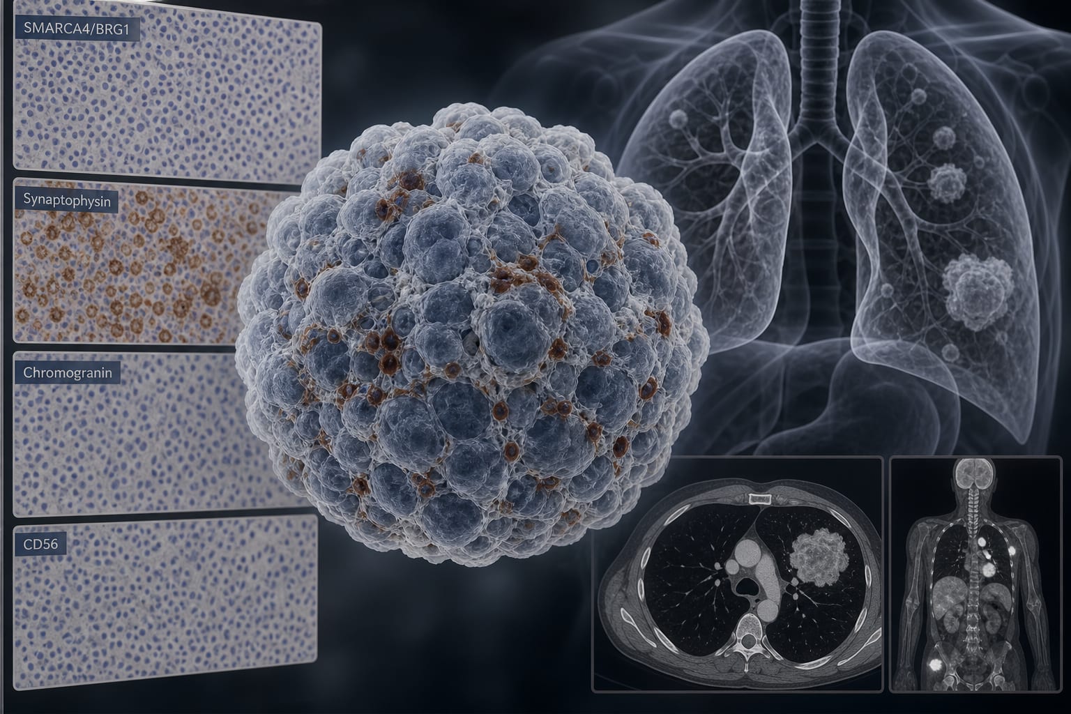
This report summarizes findings from a collection of case studies highlighting the role of PET-CT imaging in diagnosing rare neoplasms and atypical disease presentations. The studies demonstrate PET-CT's utility in staging, identifying biopsy sites, and evaluating treatment responses.
Background
The integration of PET-CT imaging in clinical practice is essential for accurate diagnosis and management of complex cases, particularly when conventional imaging methods are inconclusive.
Data Highlights
No numerical data provided in the source material.
Key Findings
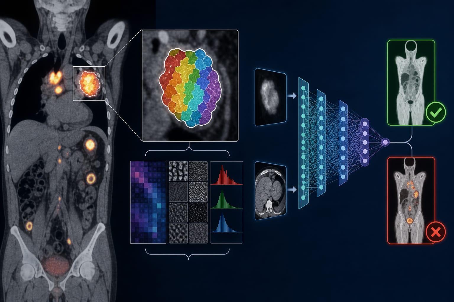
PET-CT is crucial for staging and identifying unexpected disease sites in rare neoplasms.
In plasmacytoid dendritic cell leukemia, higher SUVmax values correlate with poorer survival outcomes.
Intravenous furosemide can reduce background activity in bladder imaging, enhancing lesion visualization.
PET-CT aids in distinguishing between local recurrence and distant metastasis in previously treated cancer patients.
Case studies illustrate the importance of context-specific interpretation of PET-CT findings.
Clinical Implications
Clinicians should consider PET-CT imaging when conventional imaging fails to provide clear diagnostic information, particularly in cases of rare malignancies.
Conclusion
The findings from these case studies underscore the vital role of PET-CT in enhancing diagnostic accuracy and guiding treatment decisions in complex clinical scenarios.