Recognizing the signs of corneal ectasia can help with prevention of vision loss, and may decrease the emotional impact and functional deficits that can follow.
Specialty contact lenses are crucial in managing corneal ectasia, particularly keratoconus, which typically affects younger patients. Advances in corneal imaging and treatment options like corneal cross-linking (CXL) allow for earlier diagnosis and intervention.
Background
Keratoconus is a progressive condition that leads to corneal ectasia, often diagnosed in young adults. Early detection is essential to prevent significant visual impairment. Recent advancements in imaging technology and treatment options have improved the ability to diagnose and manage this condition effectively.
Data Highlights
Patient Age
Recommended Follow-Up
<20 years old
4-6 weeks
20-30 years old
3 months
30-40 years old
6 months
>40 years old
Annually
Key Findings
Keratoconus is typically diagnosed in patients during their second or third decade of life.
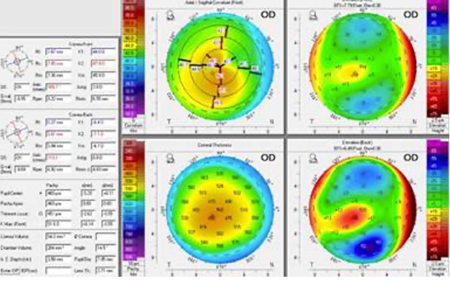
Corneal imaging technologies, including tomography and topography, are vital for early diagnosis and monitoring of keratoconus.
Signs of corneal ectasia include apical thinning, Vogt striae, and Fleisher ring.
Corneal cross-linking (CXL) can prevent progression of keratoconus and is an important treatment option.
Specialty contact lenses are effective in managing corneal ectasia, with or without vision loss.
Clinical Implications
Clinicians should prioritize early screening for keratoconus using advanced imaging techniques.
Conclusion
Early diagnosis and intervention are critical in managing keratoconus.
Bernie Iliakis, president and CEO of CorneaGen, discusses the growing momentum of CTAK to treat keratoconus, how the company is meeting the growing demand for DMEK and DSAEK tissue processing, and more.