Evaluation of intraoperative color Doppler ultrasonography in the surgical evacuation of acute intracranial hematoma: a single-center study from Botswana - Report - MDSpire
Advertisement
Evaluation of intraoperative color Doppler ultrasonography in the surgical evacuation of acute intracranial hematoma: a single-center study from Botswana
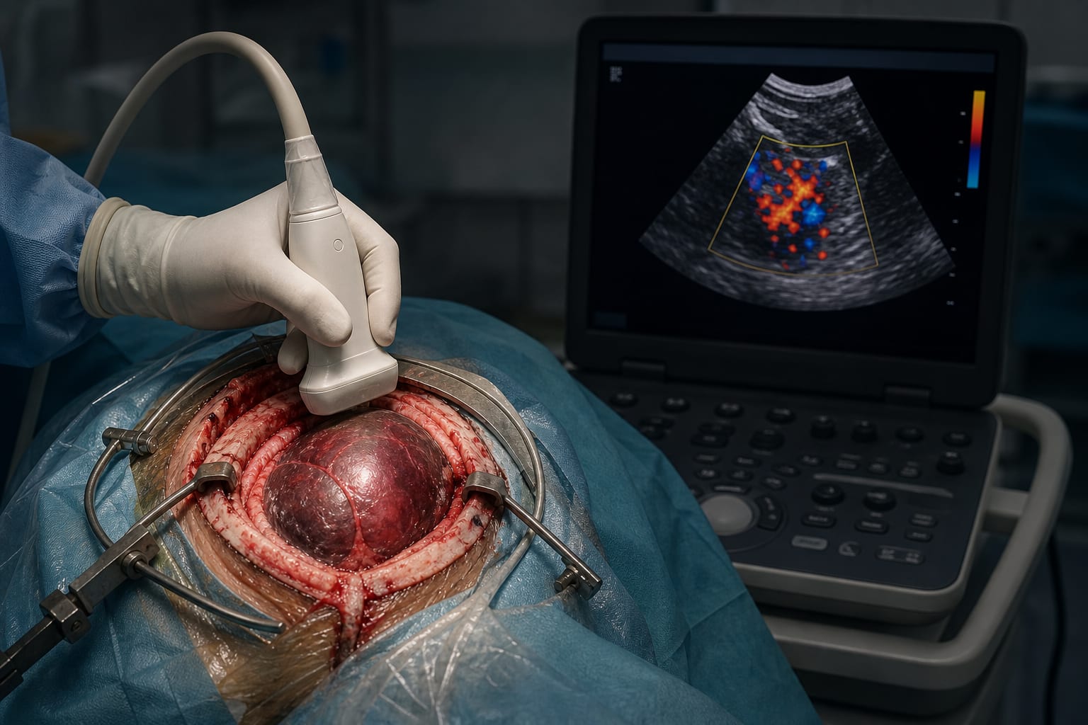
Clinical Report: Assessment of Intraoperative Color Doppler Ultrasound for Hematomas
Overview
Intraoperative color Doppler ultrasonography significantly improves hematoma clearance and neurological outcomes in patients undergoing craniotomy for acute intracranial hemorrhage. This technique is particularly beneficial in resource-limited settings, enhancing surgical precision and patient recovery.
Background
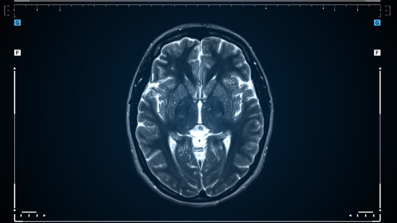
Acute intracranial hemorrhage is a critical condition requiring prompt surgical intervention to prevent neurological deterioration. Traditional imaging methods like CT scans provide static information, which may not reflect the dynamic changes occurring during surgery. The use of intraoperative color Doppler ultrasonography offers real-time insights, potentially improving surgical outcomes.
Data Highlights
Outcome
Ultrasound Group (n=34)
Control Group (n=23)
P-value
Hematoma clearance >90%
70.6%
26.1%
< 0.001
GCS Score (2 weeks post-op)
12 (10–12)
9 (8–10)
< 0.001
NIHSS Score (2 weeks post-op)
8.38 ± 2.15
11.78 ± 2.15
< 0.001
Length of hospital stay (days)
12.47 ± 5.34
16.39 ± 7.38
0.024
Key Findings
Ultrasound-guided surgery resulted in a significantly higher rate of >90% hematoma clearance (70.6% vs. 26.1%, P < 0.001).
Patients in the ultrasound group had better neurological recovery, with higher GCS scores (12 vs. 9, P < 0.001).
The NIHSS scores were lower in the ultrasound group (8.38 vs. 11.78, P < 0.001).
Hospital stays were shorter for the ultrasound group (12.47 days vs. 16.39 days, P = 0.024).
Intraoperative ultrasonography detected residual and newly formed hematomas not visible on preoperative CT.
Clinical Implications
Intraoperative color Doppler ultrasonography can enhance surgical outcomes for patients with acute intracranial hematomas by providing real-time imaging and facilitating better decision-making during surgery. This technique is especially valuable in settings with limited access to advanced imaging technologies.
Conclusion
The integration of intraoperative color Doppler ultrasonography into surgical practice for acute intracranial hematomas can lead to improved patient outcomes and should be considered in both resource-rich and resource-limited environments.