Navigated Intraoperative Ultrasound for Brainshift Correction in Glioma Surgery
Overview
Brainshift significantly impairs neuronavigation accuracy during glioma surgery. Utilizing navigated intraoperative ultrasound (iUS) with rigid image fusion (RIF) enables real-time brainshift correction, improving localization of critical structures such as the corticospinal tract and enhancing surgical decision-making.
Background
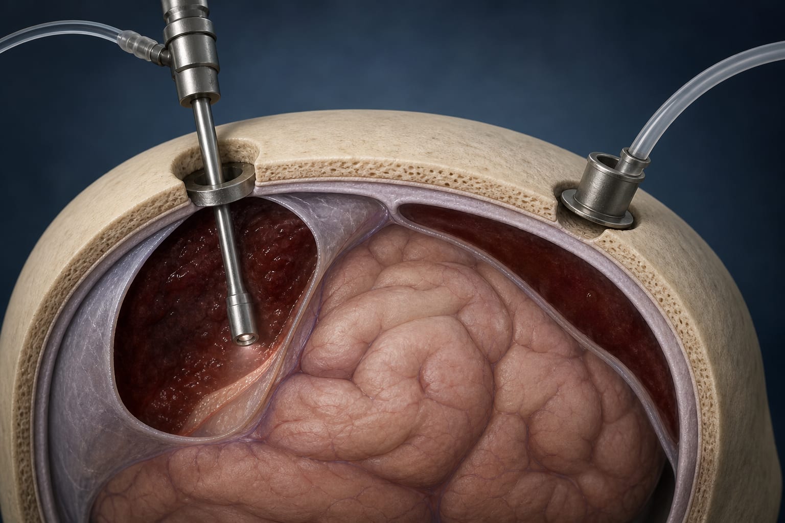
Neuronavigation relies on preoperative MRI to guide brain tumor surgeries but is limited by brainshift, a dynamic deformation of brain tissue during surgery that invalidates rigid body assumptions. Brainshift arises from factors like tissue loss, fluid shifts, and gravity, leading to inaccuracies in tumor localization and risk to eloquent brain areas. While intraoperative MRI offers precise brainshift correction, its cost and logistical challenges limit widespread use. Navigated intraoperative ultrasound, especially 3D ultrasound fused with MRI, provides a cost-effective, repeatable method to detect and correct brainshift intraoperatively.
Data Highlights
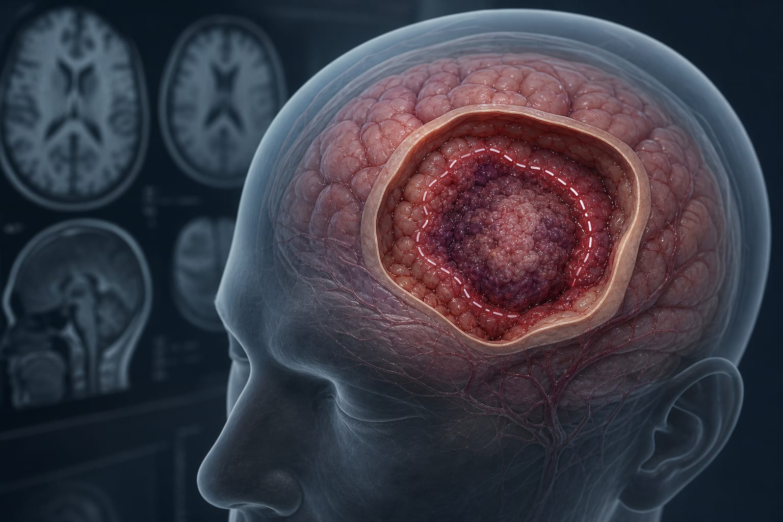
A 31-year-old female with a left frontal glioma adjacent to motor cortex and corticospinal tract underwent resection guided by navigated iUS. Serial 3D iUS scans at multiple surgical stages demonstrated brainshift occurrence and enabled correction via rigid image fusion. Post-correction imaging showed realignment of corticospinal tract location correlating with subcortical motor mapping and postoperative MRI-DTI, confirming preservation of critical pathways.
Key Findings
Brainshift occurs dynamically during glioma surgery, detectable as early as before dural opening.
Navigated 3D intraoperative ultrasound fused with preoperative MRI allows visualization and quantification of brainshift.
Corrected imaging accurately localizes the corticospinal tract at tumor margins, corroborated by intraoperative motor mapping.
Repeated brainshift correction reduces cumulative navigation errors and aids safe maximal tumor resection.
Postoperative MRI-DTI confirms preservation of eloquent white matter tracts after brainshift-corrected resection.
Clinical Implications
Incorporating navigated intraoperative ultrasound with brainshift correction enhances the accuracy of neuronavigation during glioma surgery, particularly near eloquent areas. This technique supports safer maximal tumor resection by providing real-time updates on anatomical shifts, reducing the risk of neurological deficits. Its cost-effectiveness and repeatability make it a practical adjunct to standard neuronavigation.
Conclusion
Navigated intraoperative ultrasound combined with rigid image fusion effectively compensates for brainshift during glioma surgery, improving localization of critical structures and aiding surgical decision-making. This approach enhances the safety and efficacy of tumor resection in eloquent brain regions.
References
Brainlab AG, GE Healthcare, Philips -- Navigated Intraoperative Ultrasound and MRI Fusion Techniques
Clinical Case Report -- Application of Brainshift Correction in Glioma Surgery