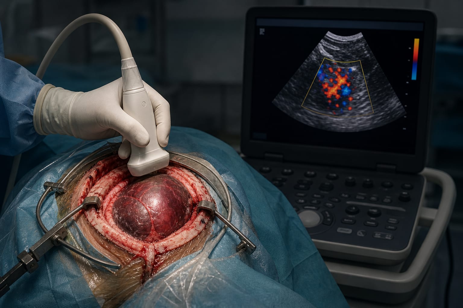
Techniques for Microsurgical Access to the Carotid Dural Rings: Stepwise Guide
Overview
This report details a meticulous step-by-step microsurgical technique for accessing the carotid dural rings, emphasizing anatomical landmarks and surgical approaches to minimize complications. It highlights the importance of careful dissection during anterior clinoidectomy and dural ring opening to avoid injury to critical neurovascular structures.
Background
The carotid dural rings, composed of proximal and distal fibrous layers, anchor the internal carotid artery (ICA) at the skull base and demarcate critical anatomical transitions. The distal dural ring (DDR) forms a tight collar around the ICA and is closely related to the optic and oculomotor nerves, as well as the cavernous sinus. Surgical access to this region is essential for managing paraclinoid aneurysms and requires precise microsurgical techniques to prevent complications such as ICA injury, aneurysm rupture, or optic nerve damage.
Data Highlights
The surgical technique involves positioning the patient prone with the head turned 30° contralaterally and fixed in a Mayfield clamp. Approaches include the conventional pterional craniotomy with a frontotemporal incision and a less invasive sphenoid ridge keyhole approach with a 5 cm curvilinear incision. Anterior clinoidectomy is performed using diamond burrs and micro-rongeurs to expose the carotid dural rings. Hemostasis in the cavernous sinus is managed with fibrin glue or hemostatic sponges. The dural rings are incised carefully under microscopic magnification to mobilize the ICA and expose aneurysm necks.
Key Findings
The carotid dural rings consist of two concentric fibrous layers anchoring the ICA at the skull base, with the distal dural ring forming a critical collar around the ICA.
The clinoid segment of the ICA lacks an external adventitial layer, increasing vulnerability during dissection.
The conventional pterional and sphenoid ridge keyhole approaches provide access to the anterior clinoid process and carotid dural rings with differing degrees of invasiveness.
Anterior clinoidectomy involves drilling the greater sphenoid wing, coagulating and incising the meningo-orbital band, and removing the anterior clinoid process to expose the carotid dural rings.
Careful dural incision along the falciform ligament and optic sheath allows mobilization of the optic nerve and exposure of the distal dural ring for safe aneurysm clipping.
Minor cavernous sinus bleeding can be controlled effectively with fibrin glue or hemostatic sponges without compromising neurovascular structures.
Clinical Implications
Understanding the detailed microsurgical anatomy and employing a stepwise approach to the carotid dural rings is essential to minimize risks during skull base surgery. Surgeons should carefully select the surgical approach based on lesion characteristics and patient anatomy, ensuring meticulous dissection to protect the ICA and cranial nerves. Effective hemostasis techniques are critical to managing cavernous sinus bleeding without causing additional injury.
Conclusion
A thorough knowledge of the carotid dural ring anatomy combined with precise microsurgical techniques facilitates safe access to the paraclinoid region. This stepwise approach reduces the risk of complications and improves surgical outcomes in complex skull base procedures.
References
Kobayashi et al. 1989 -- Description of the carotid cave