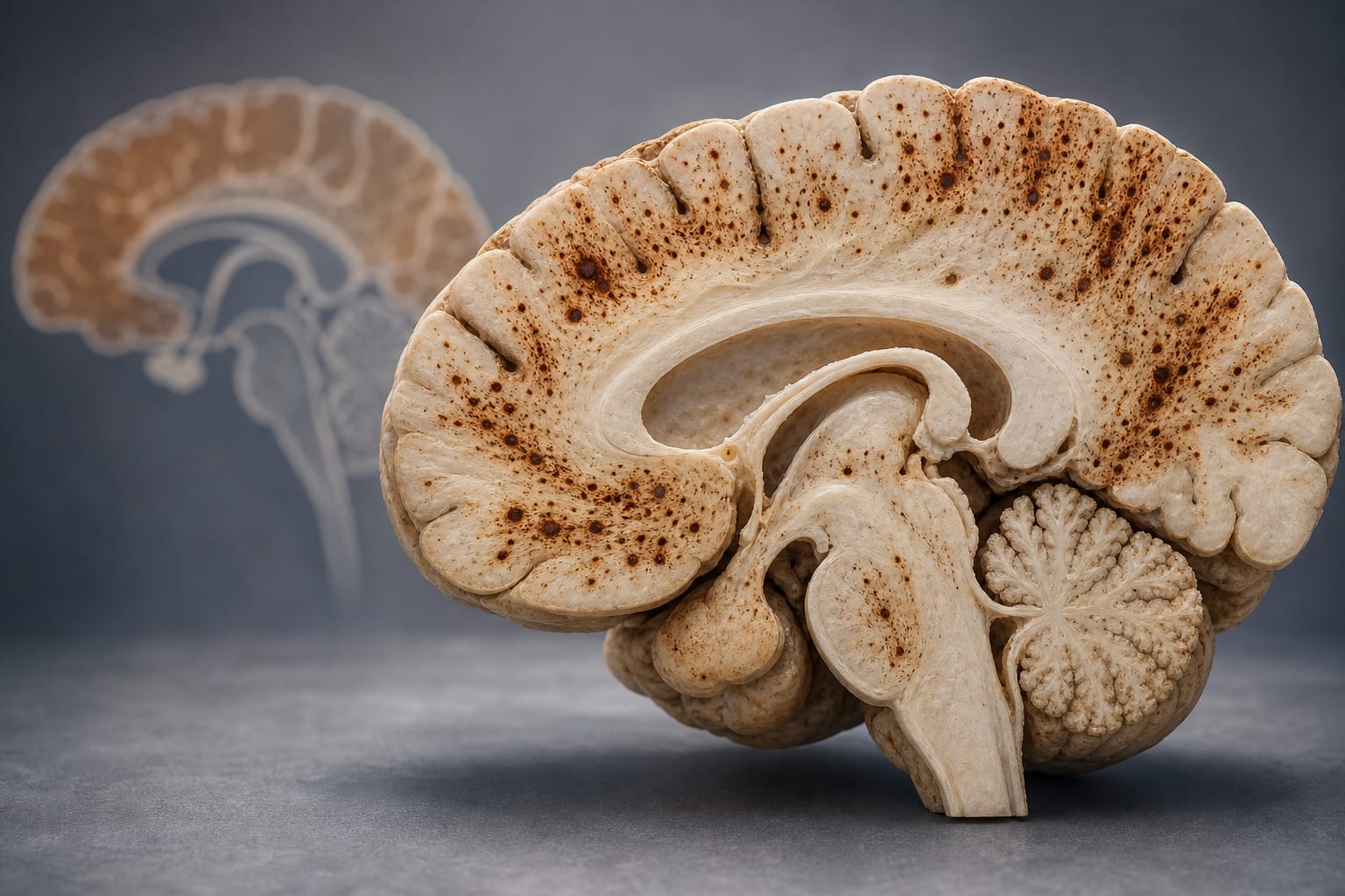
White matter changes in Alzheimer's disease: contributor, consequence or co-pathology?
-
By
-
Julia Neitzel
-
October 30, 2025
-
0 min
White Matter Alterations in Alzheimer's Disease: Cause, Effect, or Coexisting Condition?
Overview
Recent research indicates that cortical tau deposition precedes and parallels white matter degeneration in Alzheimer's disease (AD), suggesting white matter changes are a downstream consequence rather than an independent co-pathology. This challenges traditional views and highlights the potential role of tau-mediated axonal damage in early AD pathogenesis.
Background
Alzheimer's disease has traditionally been viewed as a grey matter disorder characterized by amyloid plaques and tau tangles. However, emerging evidence reveals significant white matter micro- and macrostructural changes in AD, influencing symptom onset and progression. The etiology of these white matter alterations remains debated, with some considering them independent co-pathologies linked to cerebral small vessel disease, while others propose they are integral to AD pathogenesis, possibly driven by tau and amyloid-related mechanisms.
Data Highlights
| Observation | Finding |
|---|---|
| Baseline cortical tau deposition | Associated with lower baseline volume of connected white matter regions and accelerated white matter volume loss over time |
| Baseline white matter volume | Not associated with faster tau accumulation in adjacent cortical regions |
| Rate of tau accumulation | Linked to faster degeneration of connected white matter regions |
| Participants | 726 total; 283 with longitudinal PET and MRI data from ADNI and A4/LEARN cohorts |
Key Findings
- Cortical tau deposition precedes and predicts white matter volume loss in connected brain regions.
- White matter volume at baseline does not predict subsequent tau accumulation, supporting tau-to-white-matter directionality.
- Faster tau accumulation correlates with accelerated degeneration of connected white matter tracts.
- Early tau-mediated axonal damage may occur before neuronal loss, contributing to early cognitive symptoms.
- White matter changes in AD predominantly affect limbic and temporal-parietal regions, differing from vascular pathology patterns.
- Methodological limitations include reliance on macroscopic MRI measures and white matter atlases from healthy young adults, potentially overlooking microstructural changes and individual variability.
Clinical Implications
These findings suggest that white matter alterations in AD are likely downstream effects of tau pathology rather than independent co-pathologies, emphasizing the importance of tau-targeted interventions. Differentiating AD-related white matter damage from vascular causes may be aided by assessing spatial distribution patterns on imaging. Incorporating white matter pathology into diagnostic and therapeutic models could improve early detection and treatment strategies.
Conclusion
The study supports a model where cortical tau aggregation drives connected white matter degeneration in Alzheimer's disease, highlighting white matter pathology as an integral component of AD progression rather than a separate coexisting condition.
References
This content is an AI-generated, fully rewritten summary based on a published scholarly article. It does not reproduce the original text and is not a substitute for the original publication. Readers are encouraged to consult the source for full context, data, and methodology.