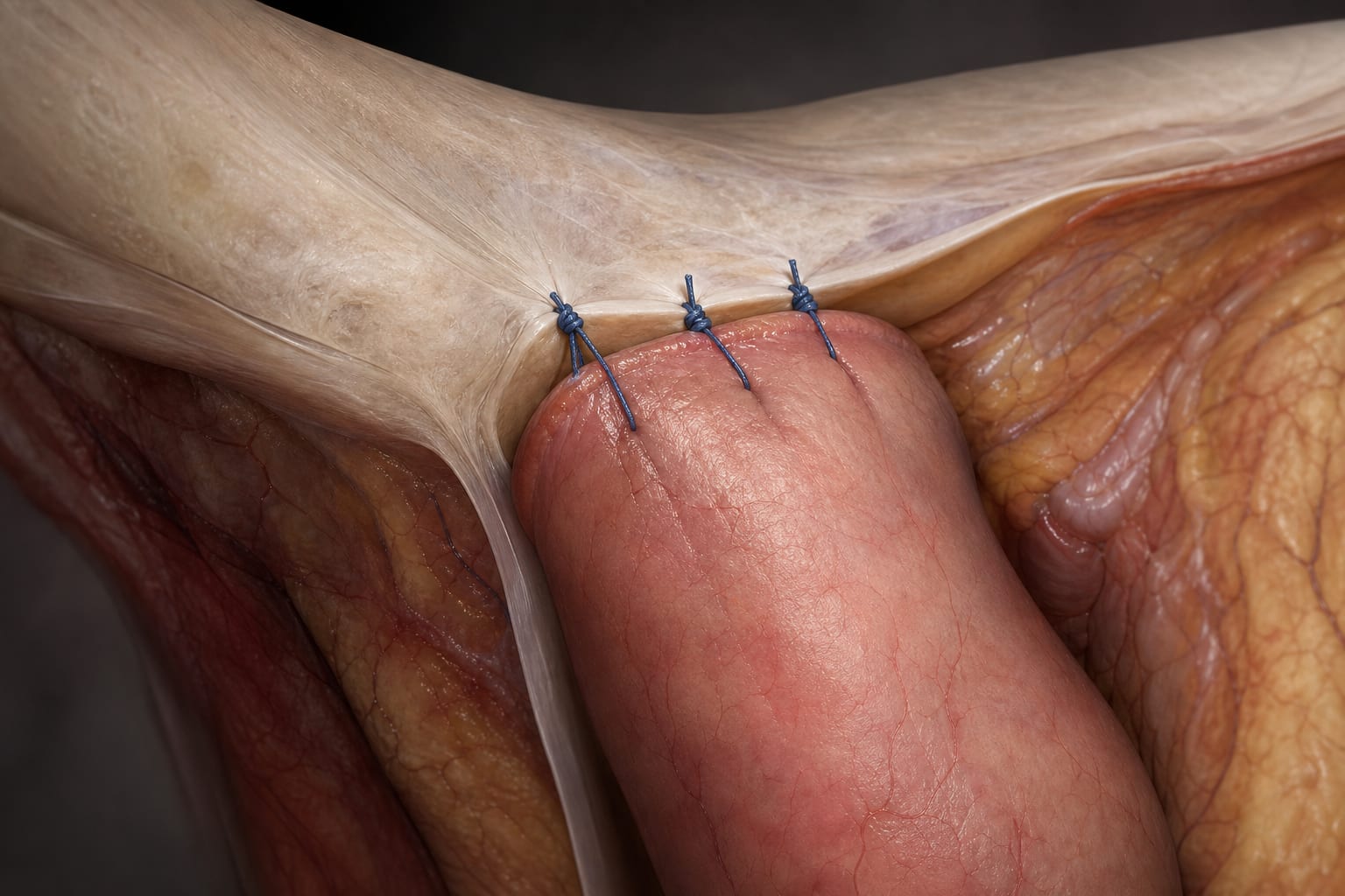
Development of Force-Measuring Apparatus for Colorectal Compression Anastomosis
Overview
This study presents the development of a novel force-measuring system designed to assess tissue penetration forces during colorectal compression anastomosis. Initial ex-vivo experiments using porcine tissue demonstrated the impact of implant design and insertion parameters on puncture forces, providing critical biomechanical data for endoscopic device development.
Background
Colorectal resection, performed for conditions such as colorectal cancer and inflammatory diseases, often requires reconnection of bowel endings via anastomosis, a step that currently limits minimally invasive approaches. Compression-based implants that pierce bowel tissue offer a promising method for secure anastomosis but require precise biomechanical characterization to optimize design and function. Porcine tissue, closely resembling human gastrointestinal anatomy and mechanics, serves as an appropriate model for these investigations. Prior studies have measured tissue puncture forces but lacked comprehensive data for multi-point compression devices needed in endoluminal applications.
Data Highlights
The experimental setup included two tissue fixation dummies spaced 4 mm apart, a disc-shaped implant dummy with symmetrically arranged spikes, and a calibrated force gauge to measure puncture forces. Previous literature reports puncture forces ranging from 0.056 N for single-needle colon piercing to 13.5 ± 3.7 N for porcine large bowel perforation. Stapling devices exert forces up to 1885 N at the stapler head, but these data are not directly applicable to multi-point compression implants. The study focused on variations in fixation point arrangements, number of implant tips, insertion speed, and acceleration to evaluate their effects on insertion force and process dynamics.
Key Findings
A novel test rig was successfully developed to reliably measure tissue penetration forces relevant to colorectal compression anastomosis.
Porcine colon tissue was validated as a suitable model due to its anatomical and biomechanical similarity to human colon.
Implant design parameters, including the number and arrangement of piercing tips, significantly influenced puncture forces.
Insertion speed and acceleration affected the force profile during tissue penetration, highlighting the importance of dynamic factors in device design.
Multi-point compression devices require biomechanical data distinct from single-needle puncture studies to ensure safe and effective tissue fixation.
Clinical Implications
The biomechanical insights gained from this study inform the design of endoscopic compression implants for colorectal anastomosis, potentially enabling less invasive and more secure reconnection of bowel segments. Understanding the force requirements and tissue interactions can improve device safety and efficacy, reducing trauma and enhancing healing outcomes. These findings support the advancement of minimally invasive colorectal surgery techniques.
Conclusion
This study provides foundational biomechanical data and a novel measurement apparatus critical for the development of compression-based colorectal anastomosis devices. The results underscore the importance of implant design and insertion dynamics in achieving secure tissue fixation with minimal trauma.
References
Heijnsdijk et al. 2005 -- Perforation forces for porcine large bowel
Kwon et al. -- Biomaterial property measurement system (BMPM) for gastrointestinal tissues
These 10 states make it more practical for physicians to participate in hospital ownership by aligning statutory structure, corporate practice of medicine rules, and population trends.