Clinical Report: Comprehensive Analysis of Diabetic Retinopathy Using UWF-OCTA
Background
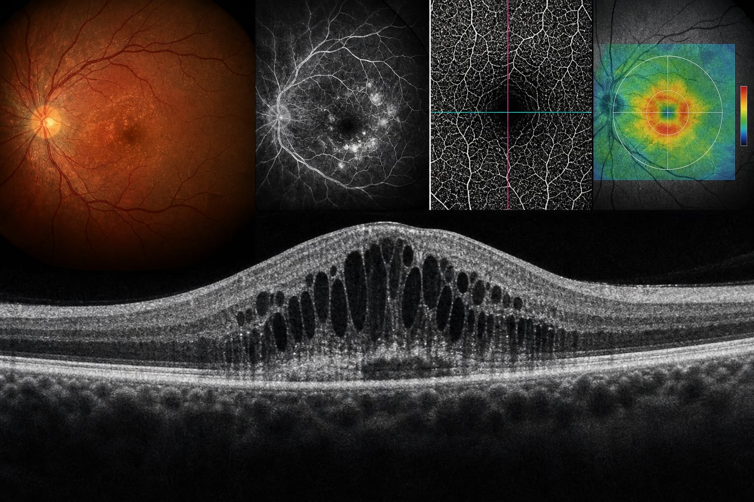
Diabetic retinopathy is a leading cause of visual impairment among working-age individuals, affecting approximately one-third of those with diabetes. Traditional diagnostic methods, such as fluorescein angiography, have limitations including invasiveness and lack of depth-resolved information. The development of UWF-OCTA offers a promising non-invasive imaging technique that enhances the visualization of retinal microvascular changes associated with DR.
Data Highlights
No numerical data provided in the source material.
Key Findings
UWF-OCTA effectively identifies and characterizes key retinal microvascular lesions, including neovascularization, non-perfusion areas, microaneurysms, and intraretinal microvascular abnormalities.
UWF-OCTA provides depth-stratified vascular visualization, surpassing the capabilities of conventional fluorescein angiography.
The technique shows high sensitivity and specificity for detecting neovascularization compared to traditional methods.
UWF-OCTA offers unique advantages, such as non-invasiveness and detailed examination of peripheral retinal vasculature.
Limitations of UWF-OCTA include potential segmentation errors and a smaller field of view compared to some traditional imaging techniques.
Clinical Implications
UWF-OCTA may serve as a valuable tool for the diagnosis and monitoring of diabetic retinopathy, complementing existing angiographic techniques. Its non-invasive nature and ability to visualize peripheral retinal changes can enhance clinical decision-making and patient management.
Conclusion
UWF-OCTA represents a significant advancement in the imaging of diabetic retinopathy, providing detailed insights into retinal vascular health while addressing some limitations of traditional methods.