Longitudinal changes in anatomic biomarkers on optical coherence tomography angiography in diabetic retinopathy - Report - MDSpire

Longitudinal changes in anatomic biomarkers on optical coherence tomography angiography in diabetic retinopathy

  • By

  • Rachel Liu

  • Isaac Bakis

  • Megan Steinkerchner

  • Kevin Sun

  • Sapna Gangaputra

  • Stephen J. Kim

  • Lok Hin Lee

  • July 9, 2026

  • 0 min

Share

Clinical Report: Temporal Changes in Anatomic Biomarkers Observed Through OCTA

Overview

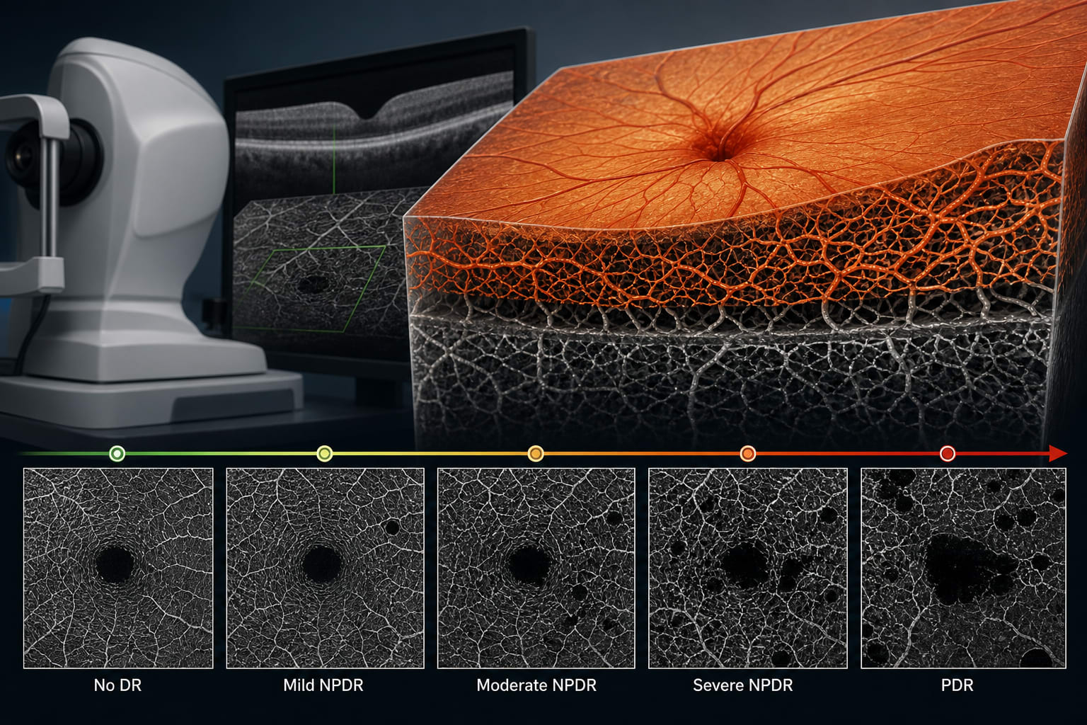
This study identifies OCTA-derived biomarkers that correlate with diabetic retinopathy (DR) severity and characterizes longitudinal retinal microvascular changes across different DR stages.

Background

Diabetic retinopathy is a leading cause of visual impairment, driven by microvascular ischemia and retinal neurodegeneration. Traditional diagnostic methods, such as fluorescein angiography, have limitations, prompting the exploration of noninvasive imaging techniques like optical coherence tomography angiography (OCTA). Understanding the dynamic changes in retinal microvasculature is crucial for timely intervention and personalized care in diabetic patients.

Data Highlights

Microvascular IndexChange per YearSeverity Group
Vessel Density-0.563 ± 0.39%Severe NPDR
Skeleton Density-0.296 ± 0.23%Severe NPDR
Acircularity Index0.16 ± 0.12%Severe NPDR vs Mild NPDR
Average Vessel Caliber-4.5e-4 ± 0.0005%Severe NPDR

Key Findings

  • Longitudinal analysis showed stage-dependent microvascular changes across DR severity groups.
  • Severe NPDR eyes exhibited greater annual declines in vessel density and skeleton density compared to controls.
  • Acircularity index increased over time, particularly in severe NPDR, indicating progressive macular ischemia.
  • Average vessel caliber decreased annually in severe NPDR compared to earlier disease stages.
  • Most OCTA metrics demonstrated small but statistically significant longitudinal trends.

Clinical Implications

OCTA-derived biomarkers can serve as noninvasive tools for monitoring DR progression.

Conclusion

The study highlights the utility of OCTA in revealing dynamic, stage-specific microvascular changes in diabetic retinopathy.

Related Resources & Content

  1. Retinal Physician, September 2021 -- OCT and OCTA in Alzheimer Disease
  2. npj Digital Medicine -- Unsupervised 3D Mapping and Analysis of Retinal Microvasculature Using RADAR
  3. Frontiers in Medicine -- Ultra-widefield optical coherence tomography angiography in diabetic retinopathy: from retinal lesions to choroidal metrics
  4. Diabetic Retinopathy Preferred Practice Pattern® - PubMed
  5. Intravitreous Aflibercept for Prevention of Vision-Threatening Complications of Diabetic Retinopathy
  6. Retinal Physician — Evolving Technology and New Terminology in Swept-Source OCT Angiography for Diabetic Retinopathy
  7. Diabetic Retinopathy Preferred Practice Pattern® - PubMed
  8. Intravitreous Aflibercept for Prevention of Vision-Threatening Complications of Diabetic Retinopathy
  9. Prognostic value of advanced imaging biomarkers for the progression of diabetic retinopathy: a systematic review | Diabetology & Metabolic Syndrome | Springer Nature Link

Original Source(s)

Related Content