Ultra-Widefield Color Fundus Photography in Diabetic Retinopathy: From Panretinal Assessment to Multimodal Integration - Report - MDSpire
Advertisement
Expanding the Scope of Diabetic Retinopathy Evaluation: The Role of Ultra-Widefield Color Fundus Photography in Comprehensive Assessment and Multimodal Approaches
Clinical Report: Expanding the Scope of Diabetic Retinopathy Evaluation
Overview
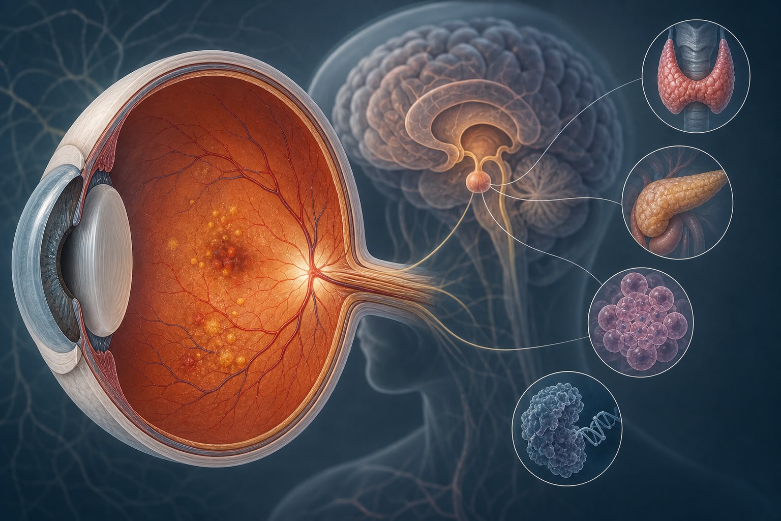
Ultra-widefield color fundus photography (UWF-CFP) significantly enhances the evaluation of diabetic retinopathy (DR) by capturing a larger retinal area, thus improving the detection of peripheral lesions. The integration of UWF-CFP with artificial intelligence and multimodal data may lead to better diagnostic accuracy and systemic risk assessment in diabetic patients.
Background
Diabetic retinopathy is a leading cause of vision loss in working-age adults, making early detection and accurate staging essential for effective management. Traditional methods, such as the ETDRS 7-field approach, often miss peripheral lesions due to limited retinal coverage. UWF-CFP addresses this limitation, offering a more comprehensive assessment of the retina.
Data Highlights
No numerical data available in the source material.
Key Findings
UWF-CFP captures up to 200° of the retina, improving visualization of peripheral lesions.
Combining UWF-CFP with AI enhances automated screening, grading, and vascular analysis.
UWF-CFP correlates with systemic complications such as diabetic nephropathy and stroke risk.
Transitioning from ETDRS 7-field to UWF-CFP can improve grading fidelity without loss of accuracy.
Peripheral lesions detected by UWF-CFP are associated with a higher risk of disease progression.
Clinical Implications
Healthcare professionals should consider incorporating UWF-CFP into routine diabetic retinopathy screenings to enhance detection of peripheral lesions. The integration of AI and multimodal imaging could lead to improved patient management and risk assessment for systemic complications.
Conclusion
The adoption of UWF-CFP represents a significant advancement in diabetic retinopathy evaluation, with the potential to transform clinical practice through enhanced detection and integrated risk assessment.