Conexiant’s news site is now MDSpire News. Learn more
Advertisement
Evaluation of Pulmonary Perfusion via Dual-Layer Dual-Energy CT for Distinguishing Acute Pulmonary Embolism from Chronic Thromboembolic Pulmonary Hypertension
Pulmonary Perfusion Assessment by Dual-Layer Dual-Energy CT Differentiates Acute PE from CTEPH
Overview
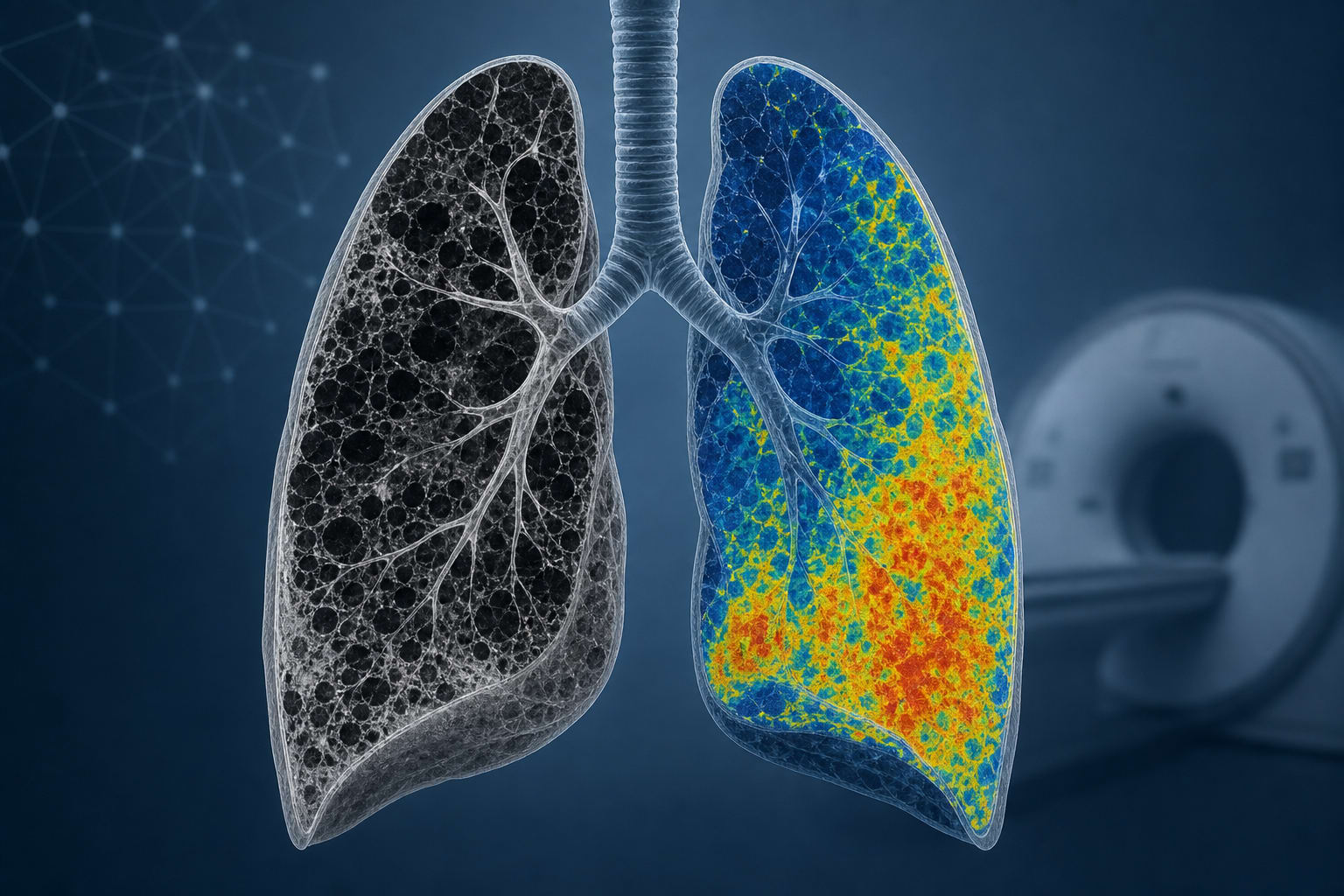
This study evaluates the use of single-phase dual-layer dual-energy CT (dlDECT) to quantify pulmonary perfusion abnormalities in acute pulmonary embolism (PE) and chronic thromboembolic pulmonary hypertension (CTEPH). The findings demonstrate that dlDECT-derived iodine density maps can distinguish acute PE from CTEPH by identifying characteristic perfusion patterns, potentially improving diagnostic accuracy.
Background
Acute PE is a common and potentially fatal cardiovascular emergency, while CTEPH is a rare but serious complication arising from unresolved or recurrent pulmonary emboli. Differentiating acute PE from CTEPH is clinically challenging due to overlapping symptoms and subtle imaging features. Conventional computed tomography pulmonary angiography (CTPA) identifies vascular and parenchymal signs but often misses subtle chronic changes. Dual-energy CT (DECT) enables visualization of pulmonary iodine distribution, serving as a surrogate for perfusion, which may help differentiate acute from chronic thromboembolic disease.
Data Highlights
The study retrospectively analyzed patients undergoing CTPA with dlDECT from 2016 to 2022, including cohorts with acute PE, CTEPH, and controls. Imaging acquisition used a 64 × 0.625 mm collimation, 120 kV tube potential, and iodine contrast bolus. Pulmonary perfusion was quantified via iodine density overlay (IDO) maps. Diagnostic criteria for CTEPH included right heart catheterization, ventilation/perfusion scintigraphy, and imaging features consistent with chronic thromboembolism after ≥3 months of anticoagulation.
Key Findings
dlDECT iodine density maps reveal distinct pulmonary perfusion patterns in acute PE versus CTEPH.
Acute PE typically shows wedge-shaped, sharply defined hypoattenuating perfusion defects corresponding to embolic occlusion.
CTEPH demonstrates patchy, multisegmental perfusion abnormalities with maintained perfusion in malperfused areas due to systemic-to-pulmonary collateral circulation.
Use of single-phase dlDECT avoids additional radiation exposure inherent to two-phase DECT protocols.
Quantitative perfusion assessment by dlDECT may reduce diagnostic delays and improve differentiation between acute and chronic thromboembolic disease.
Clinical Implications
Incorporating dlDECT iodine density mapping into routine CTPA protocols can improve diagnostic confidence in distinguishing acute PE from CTEPH, facilitating timely and appropriate management. This technique provides a noninvasive, radiation-efficient method to assess pulmonary perfusion abnormalities that are often subtle on standard imaging. Early identification of CTEPH at the index event may enable prompt referral for specialized therapies and improve patient outcomes.
Conclusion
Single-phase dual-layer dual-energy CT effectively differentiates acute pulmonary embolism from chronic thromboembolic pulmonary hypertension by characterizing distinct pulmonary perfusion patterns. This imaging modality holds promise for enhancing diagnostic accuracy and guiding clinical decision-making in thromboembolic pulmonary vascular disease.
References
ESC/ERS Guidelines 2015 -- Diagnosis and treatment of pulmonary hypertension
Philips Healthcare -- IQon Spectral CT System
Study Authors 2022 -- Evaluation of Pulmonary Perfusion via Dual-Layer Dual-Energy CT
by Roman Johannes Gertz, Felix Gerhardt, Michael Pienn, Simon Lennartz, Jan Robert Kröger, Liliana Caldeira, Lenhard Pennig, Thomas Henning Schömig, Nils Große Hokamp, David Maintz, Stephan Rosenkranz, Alexander Christian Bunck