Impact of Radiotherapy and Chemotherapy on Cerebral Volume in Brain Tumor Patients
Overview
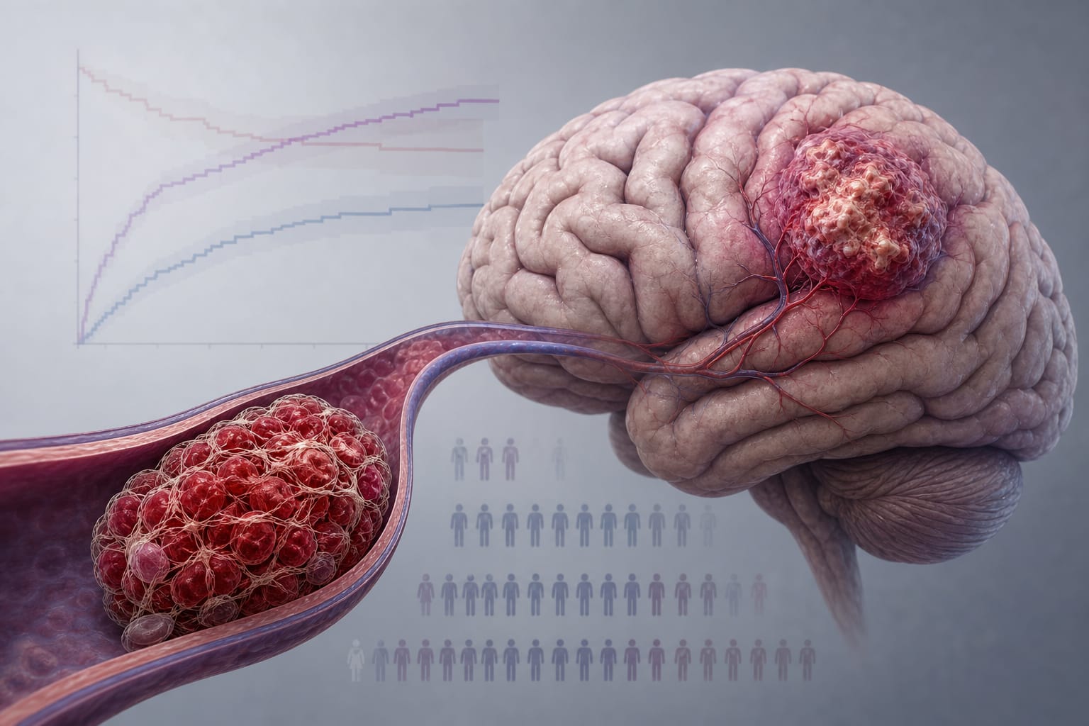
This study retrospectively analyzed brain MRI images of 47 patients with primary or metastatic brain tumors to assess changes in cerebral volumes before and after radiotherapy and/or chemotherapy. Using VolBrain and MRICloud systems, objective volumetric measurements revealed treatment-associated alterations in brain structures, contributing to understanding the effects of oncologic therapies on cerebral anatomy.
Background
Brain tumors, including gliomas, meningiomas, and metastatic lesions, cause significant morbidity and mortality and require multidisciplinary management. Treatment typically involves surgical resection supplemented by radiotherapy and chemotherapy, especially for aggressive tumors like glioblastoma multiforme. MRI volumetric analysis provides valuable anatomical and pathological information, aiding in treatment planning and prognosis. This study aimed to quantify cerebral volume changes associated with radiotherapy and chemotherapy using advanced imaging analysis tools.
Data Highlights
Parameter
Value
Number of patients included
47 (11 females, 36 males)
Age range
18–90 years
Median total radiotherapy dose
60 Gy (range 56–68 Gy)
Radiotherapy fractions
30 to 33
Patients receiving whole brain irradiation
19
Excluded patients due to missing MRI or artifacts
453 (400 no MRI, 30 no FLAIR, 23 motion artifact)
Key Findings
Volumetric MRI analysis using VolBrain and MRICloud enabled objective measurement of brain structures and tumor volumes pre- and post-treatment.
Radiotherapy doses ranged from 56 to 68 Gy, with a median of 60 Gy delivered in 30–33 fractions.
Whole brain irradiation was administered to 19 patients, potentially impacting cerebral volume more extensively.
Significant exclusion of patients due to lack of imaging or poor image quality highlights challenges in retrospective volumetric studies.
The study supports the utility of advanced imaging platforms for monitoring treatment effects on brain anatomy in oncology patients.
Clinical Implications
Quantitative volumetric assessment of brain structures before and after radiotherapy and chemotherapy can inform clinicians about treatment-related cerebral changes, potentially guiding individualized therapeutic strategies. Regular MRI follow-up with volumetric analysis may improve monitoring of tumor response and neuroanatomical preservation, ultimately enhancing patient quality of life.
Conclusion
This study demonstrates that advanced MRI volumetric tools can effectively quantify cerebral volume changes associated with radiotherapy and chemotherapy in brain tumor patients, offering valuable insights for personalized treatment planning and follow-up.
References
WHO Classification of Brain Tumors
VolBrain and MRICloud Imaging Platforms
Malatya Turgut Özal University Ethics Committee Approval 2023