Deep learning-based detection and quantification of brain metastases on black-blood imaging can provide treatment suggestions: a clinical cohort study - Report - MDSpire
Advertisement
Deep learning-based detection and quantification of brain metastases on black-blood imaging can provide treatment suggestions: a clinical cohort study
Deep Learning for Brain Metastases Detection and Treatment Guidance Using Black-Blood MRI
Overview
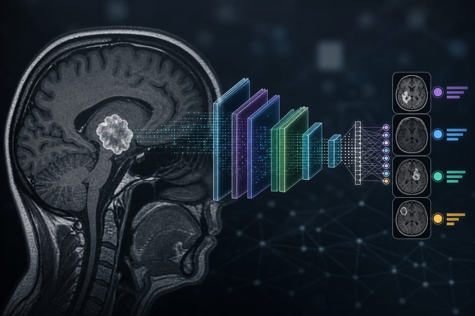
This study evaluated a deep learning system (DLS) combining gradient-echo and black-blood contrast-enhanced T1-weighted imaging for automatic detection and volumetric quantification of brain metastases (BMs). The DLS demonstrated high sensitivity and low false-positive rates, enabling treatment suggestions based on BM number and volume according to established clinical guidelines.
Background
Brain metastases are increasingly diagnosed due to improved cancer patient survival, necessitating accurate and efficient assessment for treatment planning. Traditional MRI reading often lacks segmentation and quantification of BMs, which are important for monitoring and response assessment. Deep learning systems have shown promise in automatic BM detection, but most rely on bounding box detection rather than voxel-wise segmentation for volumetry. Black-blood imaging reduces vessel-related false positives and enhances detection accuracy. Treatment decisions for BMs depend on lesion number, volume, and patient condition, with stereotactic radiosurgery (SRS), surgery, whole-brain radiotherapy (WBRT), and chemotherapy as options.
Data Highlights
Parameter
Value
Clinical cohort size
112 patients
Mean age (SD)
64.3 (9.8) years
Training dataset size
193 patients (93 with BM, 866 BMs)
DLS sensitivity for BMs <3 mm
82.4%
False-positive rate per patient
0.59
Key Findings
The DLS used paired gradient-echo and black-blood CE-T1WI images for input, improving detection accuracy.
Voxel-wise segmentation enabled volumetric quantification of BMs, facilitating treatment planning.
The DLS achieved high sensitivity (82.4%) for detecting small BMs (<3 mm) with a low false-positive rate (0.59 per patient).
Patients were stratified into treatment groups based on BM number and volume per ASCO-SNO-ASTRO and EANO-ESMO guidelines.
Group A patients with ≤2 BMs ≤5 mm diameter and volume ≤65 mm3 were recommended for imaging follow-up without immediate treatment.
The study supports the clinical utility of DLS incorporating black-blood imaging to inform treatment decisions for BM patients.
Clinical Implications
Deep learning-based automatic detection and volumetric assessment of brain metastases using black-blood MRI can streamline radiological workflows and provide objective data to guide treatment selection. Incorporating such systems may improve accuracy in identifying small lesions and support adherence to clinical guidelines for stereotactic radiosurgery and other therapies. This approach can enhance personalized treatment planning and potentially improve patient outcomes.
Conclusion
The integration of deep learning with black-blood contrast-enhanced MRI enables accurate detection and volumetric quantification of brain metastases, facilitating treatment recommendations aligned with established clinical guidelines. This technology holds promise for improving clinical decision-making in neuro-oncology.
References
ASCO-SNO-ASTRO and EANO-ESMO Practice Guidelines (2021) -- Brain Metastases Treatment Recommendations
Study on Deep Learning Detection of Brain Metastases Using Black-Blood Imaging (2023)