Clinical Report: Utilization of 3D Visualization Techniques in One-Step PTCSL
Overview
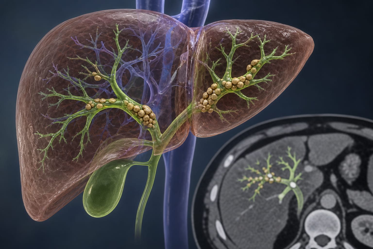
This study investigates the integration of 3D visualization technology into one-step percutaneous transhepatic cholangioscopic lithotripsy (PTCSL) for treating complex hepatolithiasis.
Background
Complex primary hepatolithiasis is a significant challenge in hepatobiliary surgery, characterized by extensive stone distribution and high recurrence rates of postoperative calculi. Traditional surgical approaches, such as hepatectomy, may not be suitable for all patients, particularly those with complex intrahepatic bile duct stones. The introduction of minimally invasive techniques, such as PTCSL, aims to improve outcomes, but challenges remain in accurately visualizing the complex anatomy involved.
Data Highlights
No numerical data or trial results were provided in the source material.
Key Findings
3D visualization technology enhances the understanding of liver anatomy and intrahepatic duct systems.
Integration of 3D imaging into PTCSL may reduce intraoperative injuries and postoperative complications.
Traditional PTCSL requires multiple surgeries and a long treatment cycle.
Patients with complex hepatolithiasis often experience high recurrence rates of stones post-surgery.
Soft choledochoscopes facilitate stone extraction compared to rigid ones.
Clinical Implications
The use of 3D visualization in PTCSL may improve surgical planning.
Conclusion
The study supports the incorporation of 3D visualization technology in the surgical management of complex hepatolithiasis.