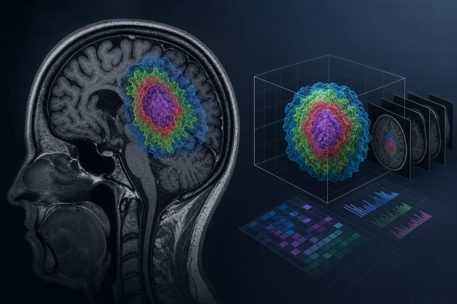
Development of the glioma imaging complexity score (GICS): a volumetric MRI-based stratification framework
-
By
-
Alex Ofori
-
Guozhu Sun
-
June 29, 2026
-
0 min