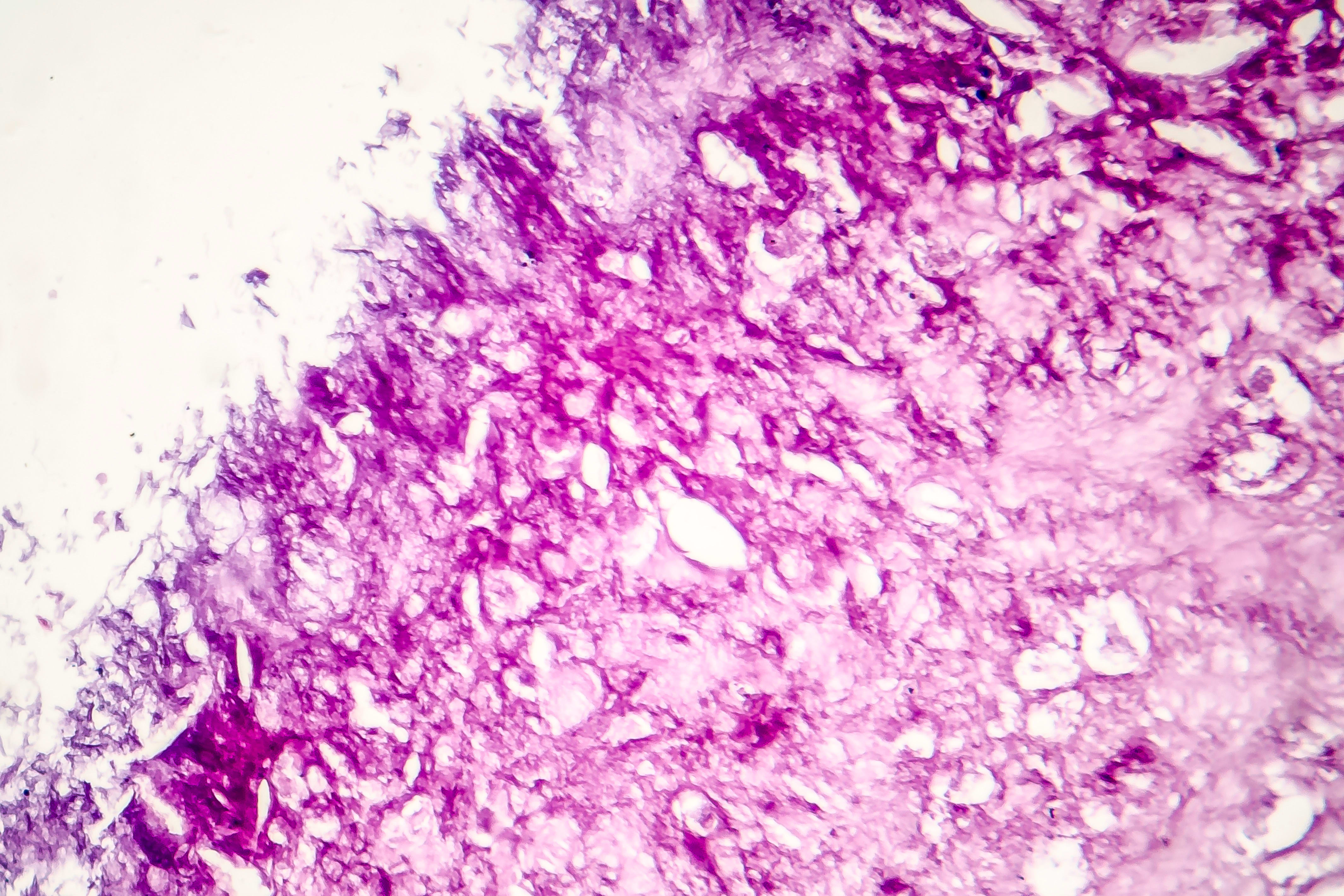
Comparative Evaluation of Radiomic Features Using Graph Neural Networks in Multi-Stained Pathological Imaging
Overview
This study evaluates the use of Radiomic features and multi-stain profiles in classifying dermatological diseases such as melanoma, eczema, and lymphoma using graph neural networks (GNNs). It demonstrates that combining graph-based modeling with Radiomic features enhances classification accuracy at both cell-wise and slide-wide levels, with dimensionality reduction techniques further improving model performance.
Background
Accurate diagnosis of dermatological conditions like melanoma, eczema, and lymphoma is challenging due to disease heterogeneity and variability in staining protocols. Traditional histopathological analysis relies heavily on expert interpretation, which can be time-consuming and prone to error. Recent advances in graph-based methods and artificial intelligence offer promising avenues to integrate biological parameters and imaging features for improved diagnostic accuracy. This study builds on prior work by exploring Radiomic features and multi-stain profiles within graph neural networks to classify pathology samples more effectively.
Data Highlights
Parameter
Value
Number of melanoma cases
20
Number of healthy tissue samples
17
Number of staining agents per sample
80–90
Image resolution
0.45 µm/pixel
Image dimensions
2018 × 2018 pixels
Key Findings
Graph neural networks effectively model pathology samples at both cell-wise and slide-wide levels for disease classification.
Radiomic features extracted from multiplex digital imaging provide valuable information for classification tasks compared to traditional multi-stain profiles.
Dimensionality reduction techniques such as UMAP and t-SNE improve classification accuracy by reducing feature complexity.
The combined use of graph structures and Radiomic features yields positive results across melanoma, eczema, and lymphoma datasets.
Expert segmentation and high-resolution imaging are critical for accurate feature extraction and graph construction.
Clinical Implications
Integrating graph neural networks with Radiomic feature analysis can enhance diagnostic accuracy in dermatology, particularly for complex diseases like melanoma, eczema, and lymphoma. This approach supports more objective and reproducible classification, potentially reducing diagnostic errors and improving patient outcomes. Additionally, dimensionality reduction methods can streamline computational workflows without compromising performance.
Conclusion
This study demonstrates that graph-based modeling combined with Radiomic features and dimensionality reduction techniques offers a robust framework for classifying multi-stained pathological images. These findings support further development of AI-assisted diagnostic tools in dermatopathology.
References
Albrecht et al. -- Feature standardization in melanoma assessment
Syrykh et al. -- Histopathological diagnosis challenges in lymphoma
Bai et al. -- AI diagnostic performance in lymphoma detection
Previous work [7,8] -- Graph-based pathology sample classification
MELC protocol [9] -- Multiplex digital imaging technique