Case report: When Behçet’s disease meets multizonal outer retinopathy and retinal pigment epitheliopathy: longitudinal multimodal imaging of an overlap phenotype - Report - MDSpire
Advertisement
Case report: When Behçet’s disease meets multizonal outer retinopathy and retinal pigment epitheliopathy: longitudinal multimodal imaging of an overlap phenotype
Clinical Report: Behçet’s Disease with Multizonal Outer Retinopathy
Overview
This case study presents a 37-year-old male with Behçet’s disease exhibiting features of multizonal outer retinopathy and retinal pigment epitheliopathy (MORR). Longitudinal multimodal imaging revealed significant retinal changes, emphasizing the need for awareness of concurrent retinal conditions in Behçet’s uveitis.
Background
Behçet’s disease is a systemic vasculitis that can lead to severe ocular complications, including uveitis and retinal vasculitis, which are major causes of vision loss. The co-occurrence of Behçet’s disease with outer retinopathies like MORR is rarely documented, highlighting a potential overlap in pathophysiology that warrants further investigation. Understanding these associations is crucial for timely diagnosis and management.
Data Highlights
No numerical data available in the article.
Key Findings
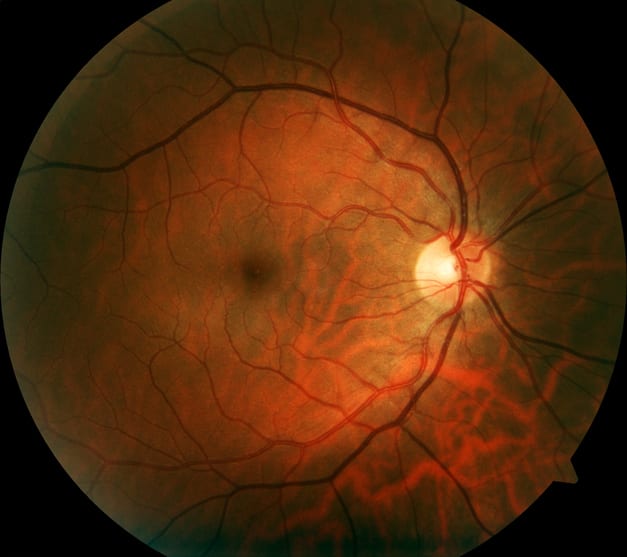
A 37-year-old male with Behçet’s disease presented with unilateral, painless vision loss.
Multimodal imaging revealed bilateral outer retinopathy and retinal vasculitis.
Best-corrected visual acuity was 20/66 in the affected eye.
Diagnosis of MORR complicated by Behçet’s uveitis was established.
Systemic treatment with corticosteroids and cyclosporine was initiated, showing favorable response.
Clinical Implications
Clinicians should be vigilant for the possibility of concurrent outer retinopathy in patients with Behçet’s uveitis, as this can complicate the clinical picture and management. The favorable response to systemic treatment in this case suggests that early intervention may be beneficial in similar presentations.
Conclusion
This case underscores the importance of multimodal imaging in diagnosing complex retinal conditions associated with Behçet’s disease. Awareness of potential overlapping features can guide effective treatment strategies.