Clinical Report: Modifying DeepLabV3+ for the Segmentation of Biopsy Lesions in Cervical Cancer
Overview
This study presents a novel approach using smartphone-assisted microscopy combined with DeepLabV3+ for the segmentation of cervical cancer lesions. The system achieved a mean Intersection over Union (IoU) of 75.8% and a Dice coefficient of 93.1%, demonstrating its potential in resource-constrained settings.
Background
Cervical cancer is a leading cause of cancer mortality, particularly in low and middle-income countries where access to advanced diagnostic tools is limited. Accurate histopathological analysis is crucial for effective diagnosis and treatment, yet many regions face significant barriers to such capabilities. This study addresses the urgent need for innovative, cost-effective solutions to improve diagnostic access in these underserved areas.
Data Highlights
Metric
Value
Mean IoU
75.8%
Dice Coefficient
93.1%
Per-Class IoU Range
74.13% - 75.41%
U-Net mIoU
56.84%
U-Net Dice
68.53%
Key Findings
The DeepLabV3+ model achieved a mean IoU of 75.8% on the validation set.
A Dice coefficient of 93.1% was recorded, indicating high segmentation accuracy.
Per-class IoU ranged from 74.13% to 75.41% across 21 histopathological feature classes.
DeepLabV3+ outperformed a U-Net baseline model under identical training conditions.
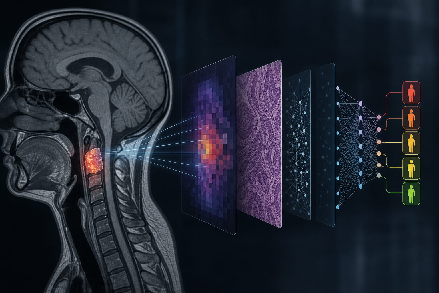
The approach integrates smartphone-assisted microscopy with advanced deep learning techniques.
Clinical Implications
The findings suggest that smartphone-assisted digital pathology can enhance diagnostic capabilities in resource-limited settings. Implementing such technology may improve early detection and treatment planning for cervical cancer, ultimately impacting patient outcomes positively.
Conclusion
This study demonstrates the feasibility of using DeepLabV3+ for effective segmentation of cervical cancer lesions in histopathological images, paving the way for improved diagnostic access in underserved regions. Further validation across multiple institutions is necessary to confirm these results.