Bone Health Deficits in Adolescent Girls with Type 1 Diabetes Before Neuropathy
Overview
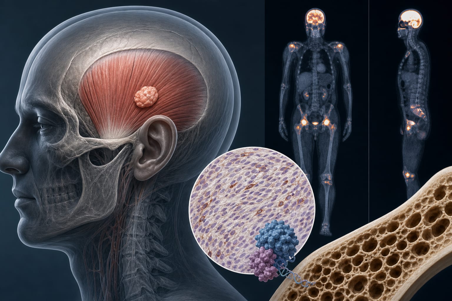
Adolescent girls with type 1 diabetes (T1D) exhibit early deficits in trabecular bone microarchitecture and suppressed bone turnover markers prior to clinical signs of peripheral neuropathy. These bone changes occur despite normal bone mineral density by DXA and are not explained by neuropathy status.
Background
Type 1 diabetes is associated with increased fracture risk beginning in childhood, yet bone health monitoring is not routinely integrated into diabetes care. Pediatric osteoporosis diagnosis relies on low bone mineral density and fracture history, but diabetic bone disease lacks distinct clinical guidelines. Diabetic peripheral neuropathy (DPN) is an early microvascular complication that correlates with bone loss in adults, but its role in early bone changes in youth with T1D remains unclear.
Data Highlights
Parameter
T1D vs Control Change
Adjusted P-value
Trabecular Bone Volume Fraction (Distal Radius)
-14.6%
0.095
Trabecular Bone Volume Fraction (Tibia)
-12.8%
0.017
Trabecular Thickness (Radius)
-8.3%
0.007
Trabecular Thickness (Tibia)
-7.5%
0.034
Cortical Bone Mineral Density (Tibia)
+8.6%
0.024
Cortical Porosity (Tibia)
-52.9%
0.012
Osteocalcin (Circulating)
-30%
0.057
Type I Collagen Cross-linked C-telopeptide
-36%
0.035
Key Findings
Adolescent girls with T1D show significant reductions in trabecular bone volume fraction and thickness at the distal radius and tibia compared to controls.
Cortical bone mineral density is increased and cortical porosity decreased in the tibia of T1D participants.
Bone turnover markers, including osteocalcin and type I collagen cross-linked C-telopeptide, are suppressed in T1D, indicating low bone formation and turnover.
Dual-energy x-ray absorptiometry (DXA) did not detect significant differences in bone mineral density between T1D and controls.
Only 9.5% of T1D participants had clinical evidence of diabetic peripheral neuropathy, which did not explain the observed bone changes.
Clinical Implications
Early bone microarchitectural deficits and suppressed bone turnover in adolescent girls with T1D occur before clinical neuropathy, suggesting that bone health deterioration begins independently of neuropathy. Routine bone health assessment using advanced imaging and biomarkers may be warranted in youth with T1D to identify early skeletal disease. Early intervention strategies, including physical activity and metabolic control, could help prevent fractures and long-term skeletal complications.
Conclusion
This study identifies early skeletal deficits in adolescent girls with T1D prior to neuropathy onset, highlighting the need for early detection and management of diabetic bone disease to reduce future fracture risk.
References
American Diabetes Association 2023 -- Standards of Medical Care in Diabetes
International Society for Clinical Densitometry 2019 -- Pediatric Osteoporosis Definition
Large Population-Based Cohort Study 2021 -- T1D and Fracture Risk Across Lifespan
Michigan Neuropathy Screening Instrument Studies 2018-2020 -- DPN Prevalence in Adolescents with Diabetes