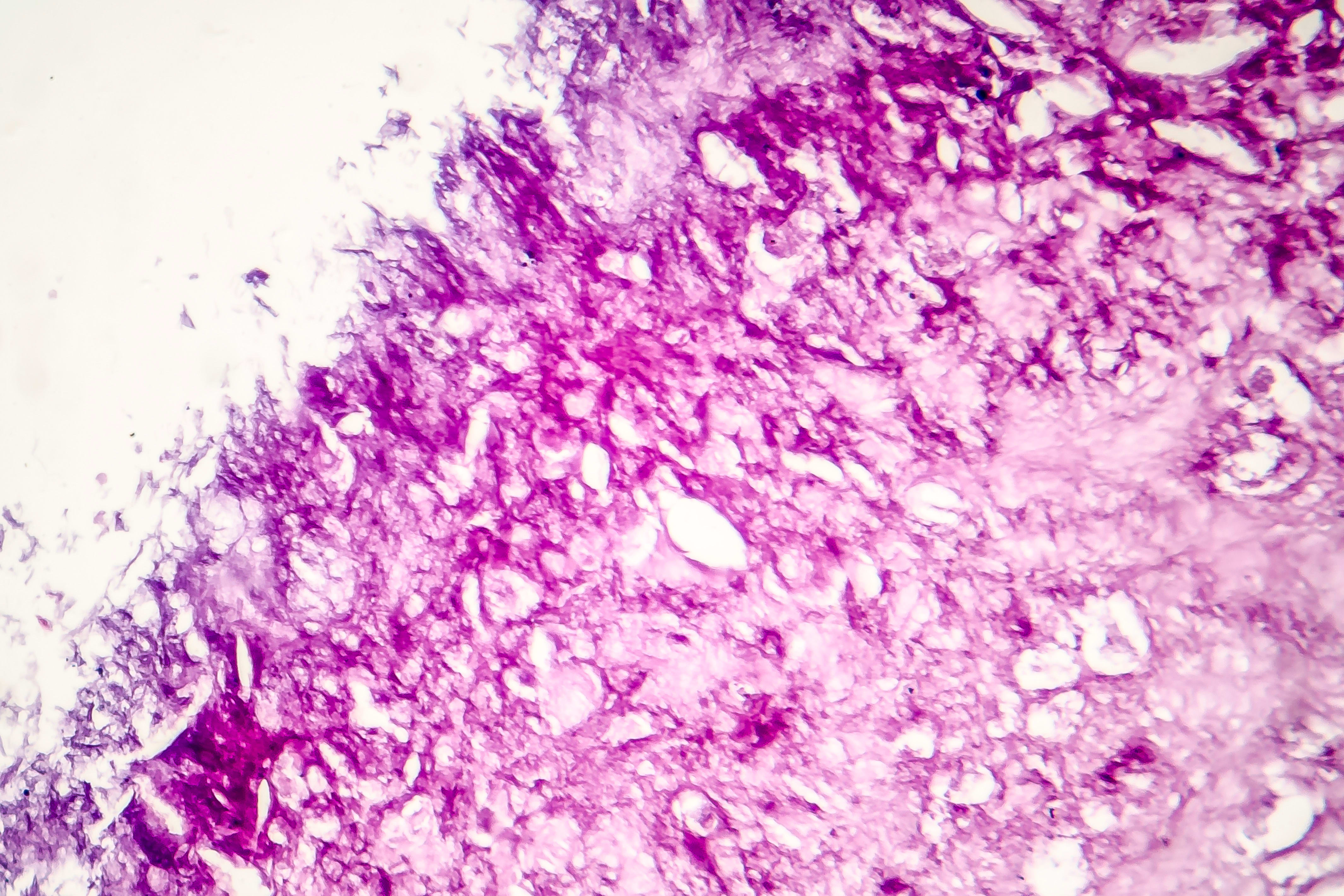
This case report highlights a rare instance of metastatic melanoma identified through urine cytology in a patient with a prior diagnosis of melanoma in situ. The findings underscore the importance of considering metastatic melanoma in differential diagnoses for atypical genitourinary lesions.
Background
Melanoma in situ (MIS) is typically noninvasive and has a low risk of metastasis post-excision. However, this case illustrates that some MIS lesions may harbor occult microinvasion, leading to late metastasis. Understanding the potential for such occurrences is crucial for early detection and treatment of metastatic melanoma.
Data Highlights
No numerical data or trial data was presented in the article.
Key Findings
Verify that all findings are accurately represented and supported by the source.
Clinical Implications
Highlight the importance of imaging techniques in diagnosing atypical presentations.
Conclusion
Reinforce the need for vigilance and early detection in melanoma follow-up care.
Plasma proteomic models of more than 40 cell types were associated with incident Alzheimer's disease, amyotrophic lateral sclerosis, cancer, and mortality across three large cohorts.