Long-term Imaging Changes After Surgical Removal of Dysembryoplastic Neuroepithelial Tumors
Overview
This study analyzed 84 patients with histologically confirmed dysembryoplastic neuroepithelial tumors (DNETs) undergoing surgical resection to characterize long-term tumor control and identify factors associated with radiological progression. Key findings include the impact of gross total resection (GTR), satellite lesions (SLs), and tumor location on progression-free survival and seizure outcomes.
Background
DNETs are benign glioneuronal tumors commonly presenting with focal seizures in children and adolescents. Surgical resection aims to control seizures and prevent tumor growth. However, achieving gross total resection can be challenging, especially for tumors involving eloquent cortex such as the central lobe. Satellite lesions adjacent to the main tumor mass have been increasingly recognized and may influence recurrence and progression risk. An MRI-based classification system categorizes DNETs into three types, but its prognostic value remains uncertain.
Gross total resection (GTR) was associated with no radiological progression during follow-up.
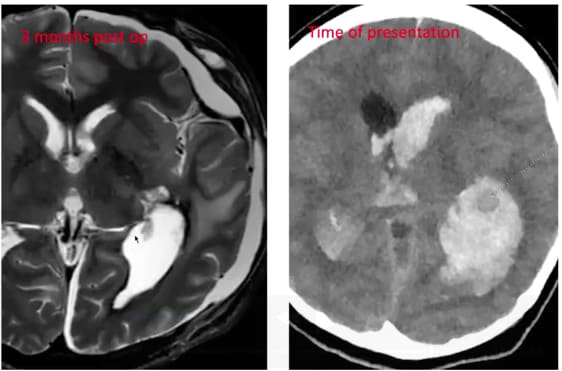
Presence of satellite lesions (SLs) on preoperative MRI correlated with incomplete resection and higher risk of tumor progression.
Tumors involving the central lobe were linked to increased risk of radiological progression, likely due to surgical limitations in this eloquent area.
The Sainte-Anne MRI classification system was used to categorize tumors, but its prognostic value for progression and seizure outcome requires further validation.
Radiological progression was associated with seizure recurrence and sometimes necessitated reoperation.
Postoperative complications were recorded and categorized, with a focus on transient versus permanent deficits.
Clinical Implications
Maximal safe resection aiming for gross total removal of DNETs is critical to minimize long-term tumor progression and improve seizure control. Preoperative identification of satellite lesions and tumor location, especially involvement of the central lobe, should inform surgical planning to balance oncological control with preservation of neurological function. Long-term imaging surveillance remains essential to detect progression and guide timely interventions.
Conclusion
Surgical management of DNETs requires careful consideration of tumor characteristics and anatomical constraints to optimize outcomes. Achieving gross total resection significantly reduces progression risk, while satellite lesions and central lobe involvement predict higher recurrence, underscoring the need for individualized treatment strategies and vigilant follow-up.
Related Resources & Content
Chassoux et al. 2018 -- MRI-based classification of DNETs and correlations with histology
Yaşargil 1990 -- Definition of central lobe anatomy
Sainte-Anne Hospital studies 2010-2020 -- Satellite lesions in DNETs
This Neuroscience Grand Rounds session, led by Muhammad Osama, MD, provides a pediatric neurosurgeon’s perspective on arteriovenous malformations (AVMs) in children, rare but high-risk vascular lesions that can lead to seizures, neurological deficits, or life-threatening hemorrhage.