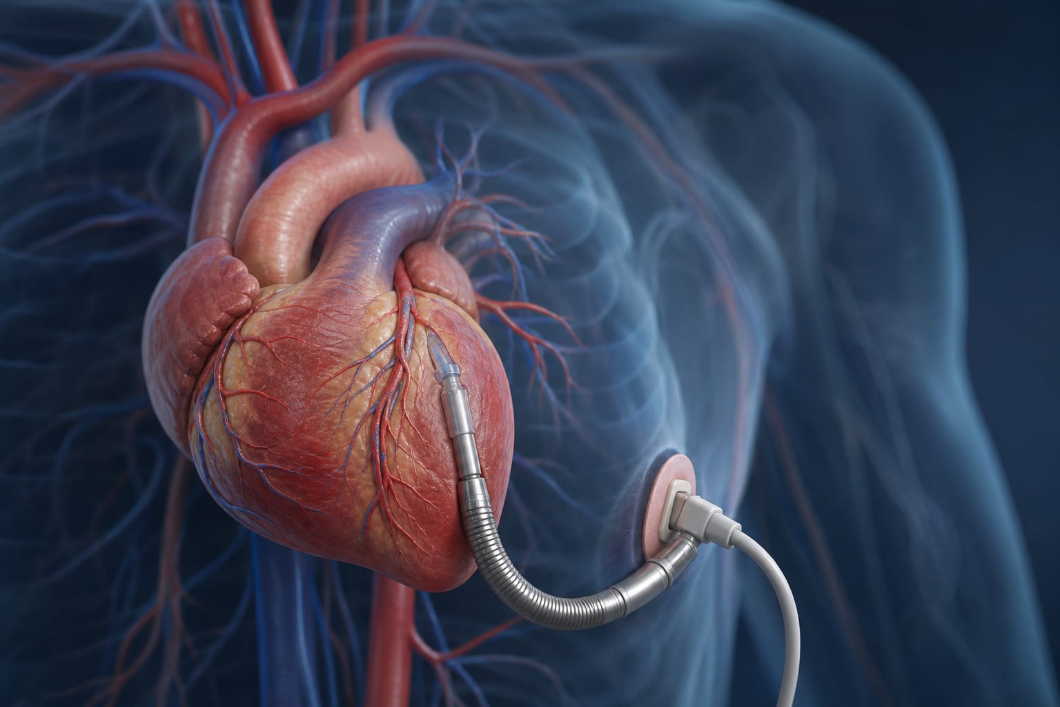
Assessment of Cardiac Function in Muscular Dystrophy via Ventriculoarterial Coupling
Overview
This study evaluated cardiovascular performance in 46 patients with muscular dystrophy (MD) using echocardiogram-derived ventriculoarterial coupling (VAC). Findings indicate that VAC is abnormal in MD patients compared to controls and correlates with disease severity and progression.
Background
Dilated cardiomyopathy is a frequent complication in adolescent and young adult patients with muscular dystrophies, often preceded by myocardial fibrosis. Traditional echocardiographic measures lack predictive power for ventricular dilation onset. Ventriculoarterial coupling (VAC), the ratio of arterial elastance to left ventricular end-systolic elastance, offers a load-adjusted index of cardiovascular efficiency and has been validated in heart failure but not previously studied in MD. This study aimed to assess VAC in MD patients and its association with disease phenotype and progression.
Data Highlights
Parameter
Value
Number of MD patients
46
Median age at diagnosis (years)
9.8 (IQR 7 - 12.2)
Median follow-up duration (years)
4.1 (IQR 3 - 6)
Duchene phenotype
35/46 (76%)
Becker phenotype
11/46 (24%)
Wheelchair use at initial echo
19/46 (41%)
Genetic mutations with high-risk cardiomyopathy
6/46 (13%)
Patients on oral steroids at initial echo
32/46 (70%)
Patients on ACEi or ARB at initial echo
30/46 (65%)
Mean LVEF at initial echo
58 ± 8%
Key Findings
VAC ratio was abnormal in MD patients compared to age-matched male controls without cardiac disease.
Higher VAC ratios correlated with worse disease phenotype, including Duchene muscular dystrophy and wheelchair dependence.
VAC worsened over longitudinal follow-up, paralleling disease progression and decline in left ventricular ejection fraction.
Patients receiving ACE inhibitors or angiotensin receptor blockers showed differences in VAC, suggesting potential therapeutic impact.
VAC provides a load-adjusted, dimensionless index reflecting ventricular-arterial interaction, superior to traditional volume-based echocardiographic measures in this population.
Clinical Implications
VAC measurement via echocardiography offers a sensitive, non-invasive tool to detect subclinical cardiac dysfunction in muscular dystrophy patients before overt ventricular dilation or heart failure. Incorporating VAC assessment may improve risk stratification and guide early therapeutic interventions, including ACEi or ARB use. Regular longitudinal VAC monitoring could help track disease progression and optimize cardiac care in this vulnerable population.
Conclusion
Echocardiogram-derived ventriculoarterial coupling is a valuable index for assessing cardiac function in muscular dystrophy, correlating with disease severity and progression. Its use may enhance early detection and management of cardiomyopathy in these patients.
References
Author/Source/Year -- Assessment of Cardiac Function in Individuals with Muscular Dystrophy