Clinical Report: Neuroimaging and Functional Changes in PCOS
Overview
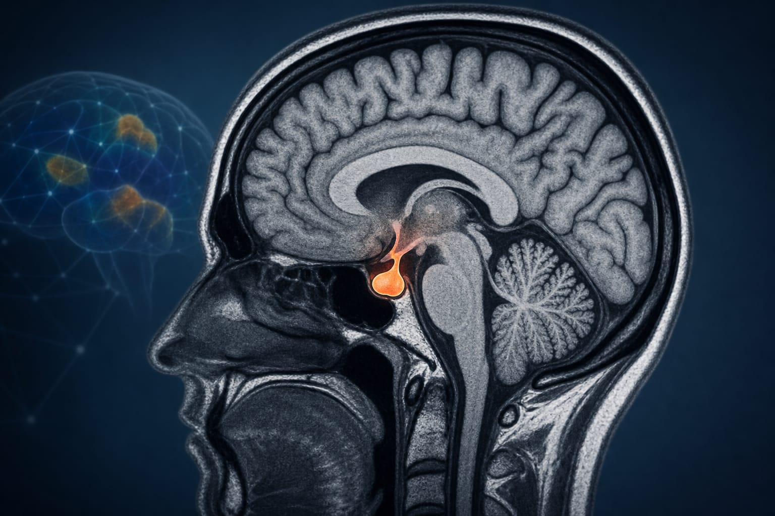
This review highlights significant neuroimaging alterations in women with polycystic ovary syndrome (PCOS), including pituitary gland enlargement and reduced gray matter volume. These changes are linked to metabolic dysregulation and cognitive-emotional disturbances, underscoring the need for further research into the pathophysiology of PCOS.
Background
Polycystic ovary syndrome (PCOS) is a prevalent endocrine disorder affecting women of reproductive age, characterized by various metabolic and reproductive dysfunctions. Understanding the neurobiological underpinnings of PCOS is crucial, as it may inform diagnostic and therapeutic strategies. Recent studies suggest that brain structural and functional changes may contribute to the clinical manifestations of PCOS, including emotional and cognitive symptoms.
Data Highlights
No specific numerical data or trial data provided in the article.
Key Findings
Enlargement of the pituitary gland observed in PCOS patients.
Reduction in gray matter volume linked to PCOS.
Damage to the corpus callosum identified in neuroimaging studies.
Altered μ-opioid receptor binding capacity in emotion-related brain regions.
Changes in activity within the right orbitofrontal cortex associated with sympathetic activation.
Structural and functional brain alterations correlated with abnormal glucose metabolism and disordered eating behaviors.
Clinical Implications
Healthcare professionals should consider the neurobiological aspects of PCOS when evaluating and treating patients. Recognizing the potential for cognitive and emotional disturbances may lead to more comprehensive management strategies, including mental health support and tailored interventions.
Conclusion
The neuroimaging findings in PCOS provide valuable insights into the disorder's pathophysiology, highlighting the interplay between endocrine, metabolic, and neurobiological factors. Further research is essential to clarify these relationships and enhance clinical outcomes.
by Ningxiao Jiang, Jie Deng, Changjin Bao, Hongmei Yin, Xianghui Zhang, Yanxia Ding, Shinan Zhang, Yingjiang Xu, Xinghua Diao, Kexin Lu, Jun Liu, Lei Han