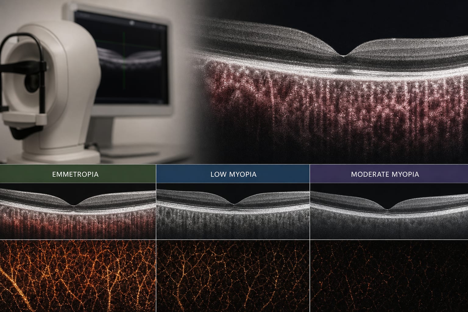
Choroidal structural and perfusion characteristics across refractive groups in children
-
By
-
Yu Liu
-
Getu Tao
-
Yifan Zhao
-
Mengyao Ma
-
Shuang Feng
-
Min Qin
-
Xiuli Bao
-
June 10, 2026
-
0 min