Clinical application study of E3D-assisted design combined with personalized 3D printed guide system in the treatment of pelvic fractures - Report - MDSpire

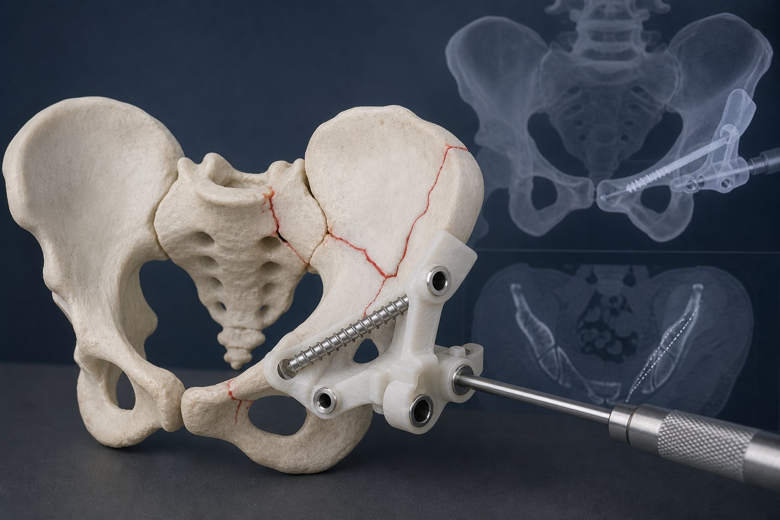
Clinical application study of E3D-assisted design combined with personalized 3D printed guide system in the treatment of pelvic fractures

  • By

  • Zheng Wang

  • JianJun Liu

  • Weidong Zhao

  • Haohui Guo

  • Jiang Wu

  • Kai Feng

  • Di Chen

  • Jun Li

  • Zhirong Chen

  • May 20, 2026

  • 0 min

Share

Clinical Report: Evaluation of E3D-Assisted Design in Pelvic Fractures

Overview

This study demonstrates that E3D-assisted design combined with personalized 3D-printed guides significantly reduces operation time and improves screw placement accuracy in pelvic fracture surgeries compared to traditional fluoroscopic methods. The findings highlight the potential for enhanced safety and efficiency in minimally invasive orthopedic procedures.

Background

Pelvic fractures, often resulting from high-energy trauma, pose significant surgical challenges due to their complexity and associated risks, including massive blood loss and damage to vital structures. Traditional surgical methods are associated with long operation times and increased risk of complications. The advent of minimally invasive techniques, particularly those utilizing 3D printing and computer-assisted design, offers promising alternatives that may improve surgical outcomes and reduce recovery times.

Data Highlights

ParameterExperimental GroupControl Groupp-value
Operation Time (min)53.05 ± 19.0481.05 ± 24.68< 0.001
Screw Placement Accuracy (tip, mm)4.63 ± 2.727.38 ± 5.920.04
Screw Placement Accuracy (pedicle, mm)6.75 ± 3.0610.15 ± 1.320.03
Screw Placement Accuracy (sacroiliac joint, mm)5.79 ± 3.388.80 ± 5.780.03

Key Findings

  • E3D-assisted design significantly reduced operation time compared to traditional methods.
  • Screw placement accuracy was higher in the experimental group across multiple measurement points.
  • No significant difference in fracture reduction quality was observed between the two groups.
  • The use of personalized 3D-printed guides enhances safety and reduces the learning curve for clinicians.
  • Minimally invasive techniques are increasingly favored due to their reduced trauma and bleeding.

Clinical Implications

The findings suggest that integrating E3D-assisted design and 3D-printed guides in surgical practice can lead to more efficient and safer procedures for pelvic fractures. Clinicians may consider adopting these technologies to improve patient outcomes and streamline surgical workflows.

Conclusion

E3D-assisted design combined with personalized 3D-printed guides represents a significant advancement in the management of pelvic fractures, offering reduced operation times and enhanced screw placement accuracy. This approach may facilitate broader adoption of minimally invasive techniques in orthopedic surgery.

Related Resources & Content

  1. Sustained Success of Customized 3D Printed Titanium Implants in Complex Pelvic Reconstruction: Insights from 106 Cases Over a Decade
  2. Next-Day Acetabular Fracture Surgery Utilizing 3D Printing with a Surface Filtering Pipeline: Feasibility Study and One-Year Clinical Outcomes
  3. Impact of Fitting Location and Design on Positioning Accuracy of Custom 3D-Printed Surgical Guides for the Radius
  4. Evaluation of Sacroiliac Screw Insertion Using 3D-Printed Templates Versus X-Ray Fluoroscopy Guidance
  5. Fractures (complex): assessment and management
  6. Fractures (complex): assessment and management
  7. Comparison of 3D Navigation and Fluoroscopy for Pelvic Fixation
  8. Frontiers | Clinical efficacy analysis of anterior superior iliac spine 3D-printed guide plate-assisted sacroiliac screw placement for the treatment of pelvic fractures

Original Source(s)

Related Content