Subcortical language localization using sign language and awake craniotomy for dominant posterior temporal glioma resection in a hearing-impaired patient - Report - MDSpire
Advertisement
Subcortical language localization using sign language and awake craniotomy for dominant posterior temporal glioma resection in a hearing-impaired patient
Localization of Language Functions in Subcortical Areas During Awake Craniotomy in Hearing-Impaired Patient
Overview
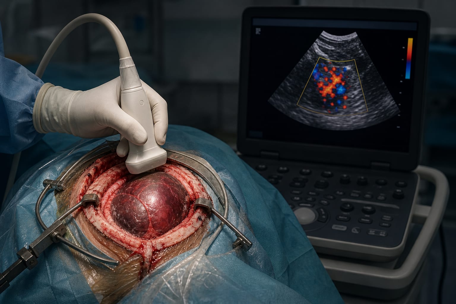
This case report describes the first awake craniotomy with direct cortical stimulation (DCS) testing both sign language and spoken language in a post-lingual deaf patient. Subcortical stimulation near the arcuate fasciculus (AF) and inferior fronto-occipital fasciculus (IFOF) elicited phonemic paraphasias, guiding the extent of tumor resection while preserving language function.
Background
Intraoperative direct cortical stimulation (DCS) is the gold standard for mapping eloquent brain areas during tumor resection to maximize removal while preserving function. Language mapping during awake craniotomy is challenging in hearing-impaired patients, with few reported cases involving sign language. This report presents a unique case of a post-lingual deaf patient proficient in American Sign Language (ASL) and spoken English undergoing awake mapping for both modalities. Preoperative functional MRI and diffusion tensor imaging (DTI) were used to localize language areas and critical white matter tracts.
Data Highlights
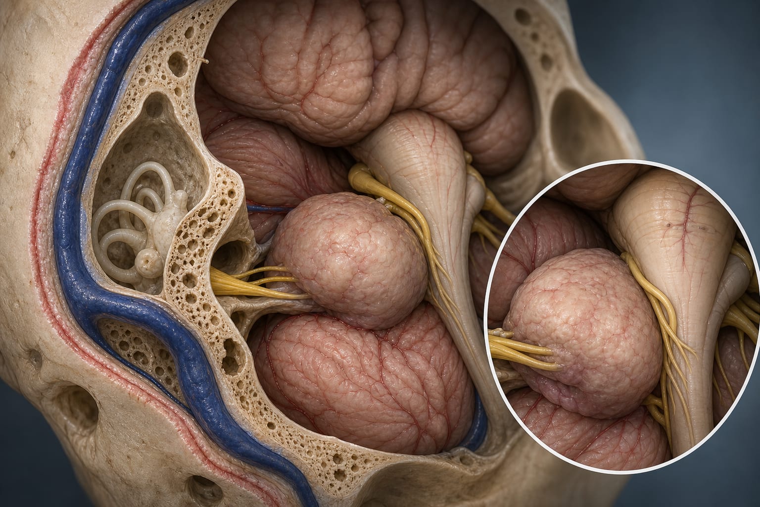
Preoperative fMRI showed left hemisphere language lateralization near the posterior temporal lesion. DTI identified the arcuate fasciculus (AF) anterior and medial to the lesion and the inferior fronto-occipital fasciculus (IFOF) inferior and medial to the lesion. Intraoperative subcortical stimulation at these tracts elicited phonemic paraphasias for both picture and ASL gesture cues, marking the medial resection boundary. Postoperative MRI confirmed gross total resection.
Key Findings
First reported awake craniotomy case testing both sign language and spoken language in a hearing-impaired patient.
Preoperative fMRI and DTI identified language lateralization and critical white matter tracts (AF and IFOF) near the tumor.
Cortical mapping was negative, but subcortical stimulation near AF and IFOF elicited phonemic paraphasias in both language modalities.
Phonemic paraphasias included substitutions, neologisms, and perseverations, indicating language function involvement.
Subcortical mapping findings guided the extent of tumor resection, preserving language function.
Postoperative imaging confirmed gross total resection without reported language deficits.
Clinical Implications
This case demonstrates the feasibility and importance of awake mapping for both sign and spoken language in hearing-impaired patients undergoing tumor resection near eloquent areas. Incorporating preoperative fMRI and DTI tractography enhances localization of critical language pathways. Subcortical stimulation can reveal language function involvement not detected by cortical mapping alone, guiding safer resection boundaries.
Conclusion
Awake craniotomy with multimodal language mapping, including sign language, is feasible and critical for preserving language function in hearing-impaired patients with dominant posterior temporal gliomas. Subcortical stimulation near AF and IFOF is essential for identifying language-related white matter tracts to maximize safe tumor resection.
References
Sabsevitz et al. -- Neuromapper Testing Platform
Synaptive Medical® -- Modus Plan™ Software
Previous Cases of DCS in Deaf Patients [3,6,11,13]