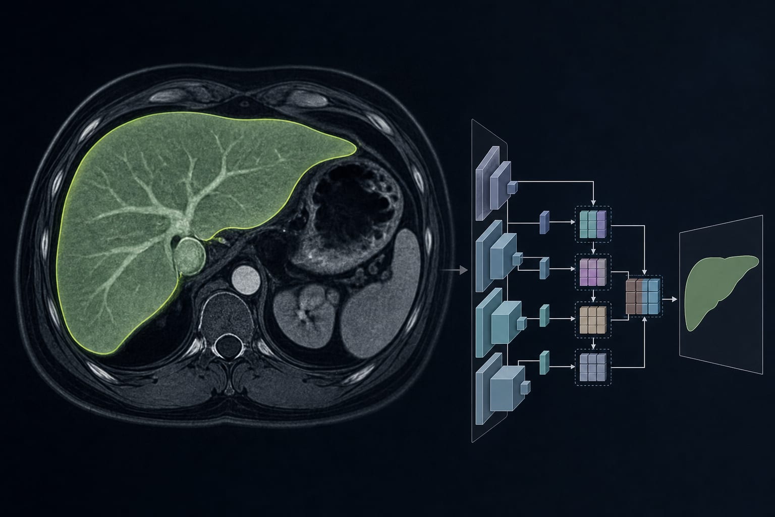
Clinical Report: A Novel Pyramid Scene Parsing Model for Liver Segmentation
Overview
The PSP-EffB0-MSFF model demonstrates high accuracy in liver segmentation from MRI scans, achieving significant performance metrics across two datasets. This automated approach addresses the challenges of manual segmentation, providing reliable and consistent results.
Background
Accurate liver segmentation is crucial for diagnosing and managing liver diseases, which are on the rise due to lifestyle changes. Manual segmentation is labor-intensive and subject to variability among operators, highlighting the need for effective automated solutions. The proposed model leverages advanced deep learning techniques to enhance segmentation accuracy in MRI imaging.
Data Highlights
Dataset
IoU
Dice Score
Hausdorff Distance
Average Symmetric Surface Distance
DLDS
0.905 ± 0.038
0.913 ± 0.09
7.31 ± 3.91
2.66 ± 3.06
CirrMRI600+
0.86 ± 0.01
0.90 ± 0.02
6.20 ± 0.60
9.80 ± 1.50
Key Findings
The PSP-EffB0-MSFF model utilizes EfficientNetB0 to reduce computational costs.
Achieved an intersection over union (IoU) of 0.905 on the DLDS dataset.
On the CirrMRI600+ dataset, the model achieved a Dice score of 0.90.
Demonstrated a Hausdorff distance of 6.20 ± 0.60 on the CirrMRI600+ dataset.
The model requires 14.91 GFLOPs for processing.
Clinical Implications
The PSP-EffB0-MSFF model can significantly enhance the efficiency of liver segmentation in clinical practice, reducing the burden of manual annotation for radiologists. Its high accuracy supports better decision-making in the management of liver diseases, including surgical planning and treatment evaluation.
Conclusion
The proposed model offers a promising automated solution for liver segmentation in MRI scans, demonstrating reliable performance across multiple datasets. This advancement could facilitate improved clinical outcomes in liver disease management.