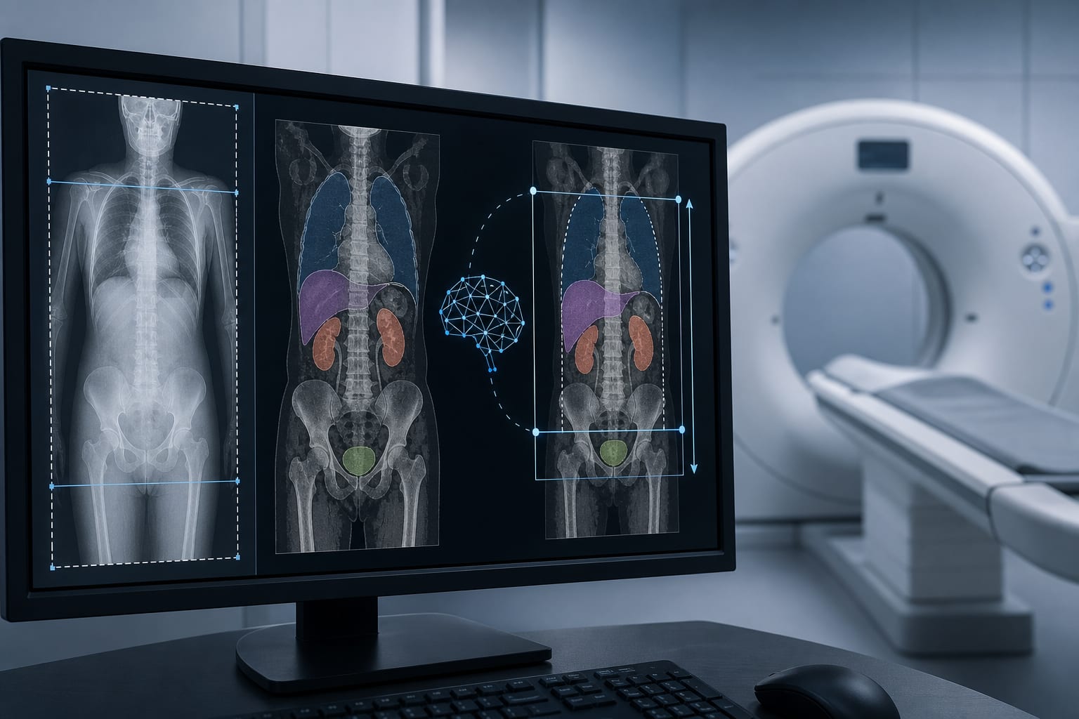
Radiopathomics Model Predicts Liver Metastases in Colorectal Cancer Patients
Overview
A novel radiopathomics model combining CT radiomics and clinical features was developed to predict postoperative liver metastases in colorectal cancer (CRC) patients. This model demonstrated improved predictive accuracy over traditional clinical or radiomics models alone.
Background
Colorectal cancer is a leading cause of cancer mortality, primarily due to postoperative liver metastases occurring in at least 50% of patients. The conventional TNM staging system lacks sufficient predictive accuracy for liver metastases, necessitating more precise tools. Pathological examination remains the gold standard but is invasive and unsuitable for frequent use. Radiomics, which extracts quantitative imaging features from CT scans, offers a noninvasive approach to characterize tumor heterogeneity and metastasis risk preoperatively.
Data Highlights
The study retrospectively enrolled CRC patients with preoperative CT scans and clinical data from 2017 to 2022. Radiomics features were extracted from manually contoured tumor regions on CT images, standardized, and selected using t-tests and L1-regularized logistic regression. Clinical features including age, sex, BMI, tumor markers (CEA, CA19-9), and tumor staging were collected. Separate clinical and radiomics models were constructed using random forest classifiers, and combined using score-level fusion strategies to enhance predictive performance.
Key Findings
The radiopathomics model integrating CT radiomics and clinical data outperformed models based on either data type alone in predicting postoperative liver metastases.
Feature selection combining statistical tests and L1 regularization effectively identified discriminative radiomics features related to tumor phenotype.
Manual tumor segmentation on CT images by experienced radiologists ensured accurate region-of-interest delineation for feature extraction.
Score-level fusion strategies (minimum, maximum, weighted) were explored to combine predictive scores from clinical and radiomics models, improving overall prediction accuracy.
The model development adhered to international biomarker standardization guidelines to ensure reproducibility and scientific rigor.
Clinical Implications
This radiopathomics model offers a noninvasive, preoperative tool to stratify CRC patients by risk of liver metastases, potentially guiding personalized treatment decisions and surveillance strategies. Incorporating quantitative imaging features with clinical data may enhance early identification of high-risk patients who could benefit from intensified therapy or closer follow-up. Adoption of such models could improve prognostication beyond traditional TNM staging.
Conclusion
The study successfully developed and validated a combined radiopathomics model that improves prediction of postoperative liver metastases in colorectal cancer patients, highlighting the value of integrating imaging biomarkers with clinical parameters for precision oncology.
References
Colorectal Cancer Statistics and Mortality -- Global Cancer Observatory
TNM Staging Limitations in CRC -- Clinical Oncology Reviews
Radiomics and Tumor Heterogeneity -- Imaging Biomarkers Journal
Radiopathomics Model Development -- Affiliated Hospital of Qingdao University Study