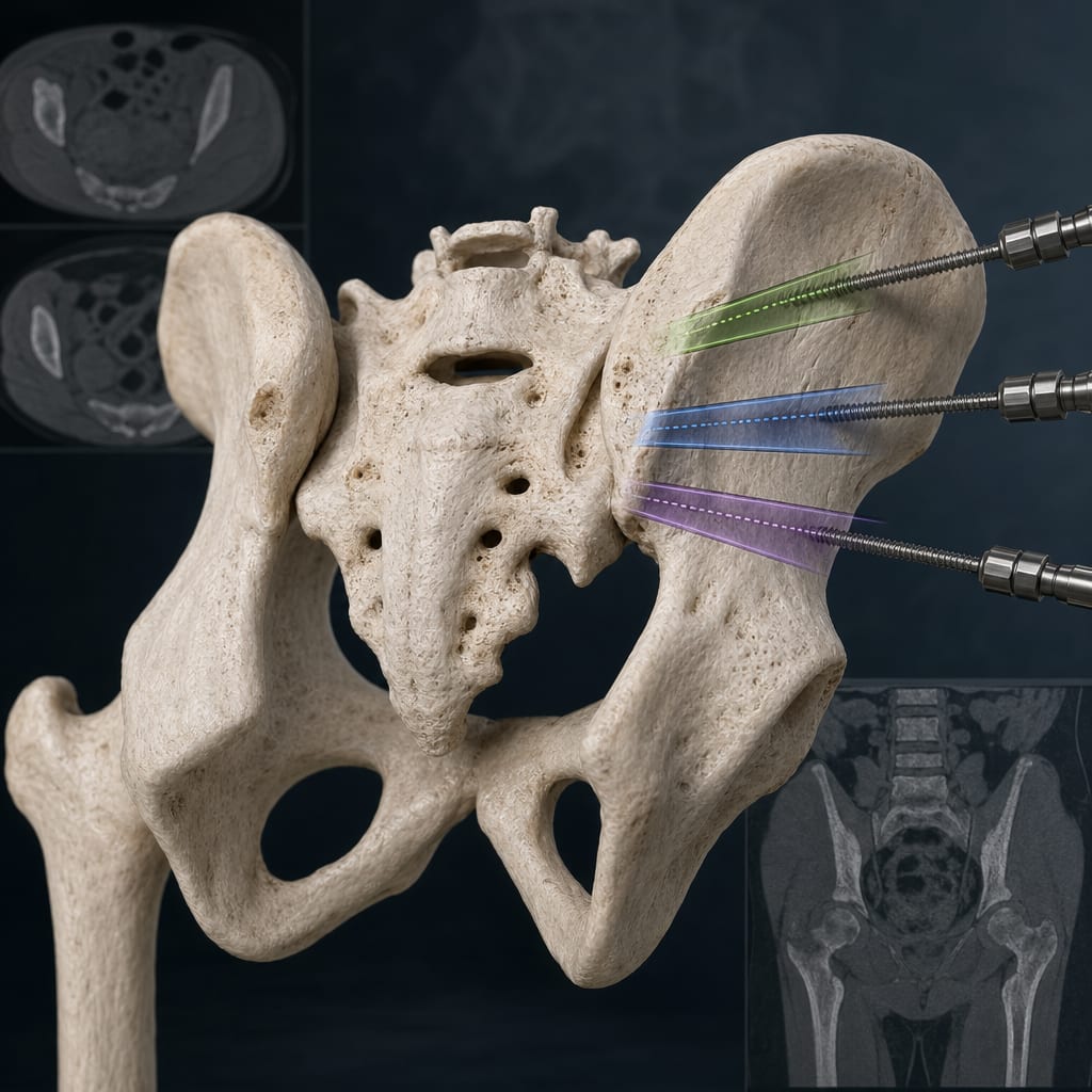
Clinical Report: Three-Dimensional CT Analysis of Safe Anatomical Pathways for Schanz Screw Insertion in Adolescent Pelvic Ring Fractures
Overview
This study identifies safe anatomical pathways for Schanz screw insertion in adolescents with unstable pelvic fractures, utilizing 3D CT imaging. The findings emphasize the importance of adhering to specific entry points and angles to minimize risks during surgical procedures.
Background
Incorporate statistics or references to substantiate the increase in adolescent pelvic fractures.
Data Highlights
Pathway
Entry Point (mm posterior to ASIS)
Horizontal Angle (°)
Sagittal Angle (°)
Iliac Wing Corridor I1
16.9–18.7
22.5–25.8
53.7–61.2
Iliac Wing Corridor I2
49.4–53.6
21.5–25.1
61.3–66.1
Supra-acetabular Corridor A
AIIS-PSIS plane
58.2–61.3
23.0–25.6
Key Findings
The iliac wing corridor transitions from a proximal rectangular shape to a distal funnel-like constriction.
Entry points for Schanz screws require precise angles to minimize perforation risks.
The supra-acetabular corridor has an S-shaped configuration with significant mid-corridor constriction.
Patient-specific preoperative planning enhances surgical safety and fixation stability.
Compliance with established entry points and angles is crucial for successful outcomes.
Clinical Implications
Surgeons should utilize 3D CT imaging for preoperative planning to ensure safe Schanz screw insertion in adolescents. Adhering to the identified entry points and angles can significantly reduce the risk of complications during external fixation procedures.
Conclusion
Highlight the study's potential influence on future research and clinical practices.