Degeneration rather than hypoplasia may be the pathologic feature of the endolymphatic sac in patients with ipsilateral delayed endolymphatic hydrops - Report - MDSpire

Pathological Characteristics of the Endolymphatic Sac in Patients with Ipsilateral Delayed Endolymphatic Hydrops: Emphasizing Degeneration Over Hypoplasia

  • By

  • Kaijun Xia

  • Ping Lei

  • Qin Liu

  • Yingzhao Liu

  • Cen Chen

  • Xiaoye Chen

  • Xingqian Shen

  • Ziying Xu

  • Bo Liu

  • Hongjun Xiao

  • Yangming Leng

  • July 21, 2026

Share

Clinical Report: Pathological Characteristics of the Endolymphatic Sac in DEH

Overview

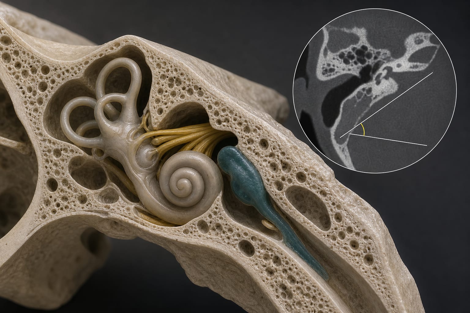
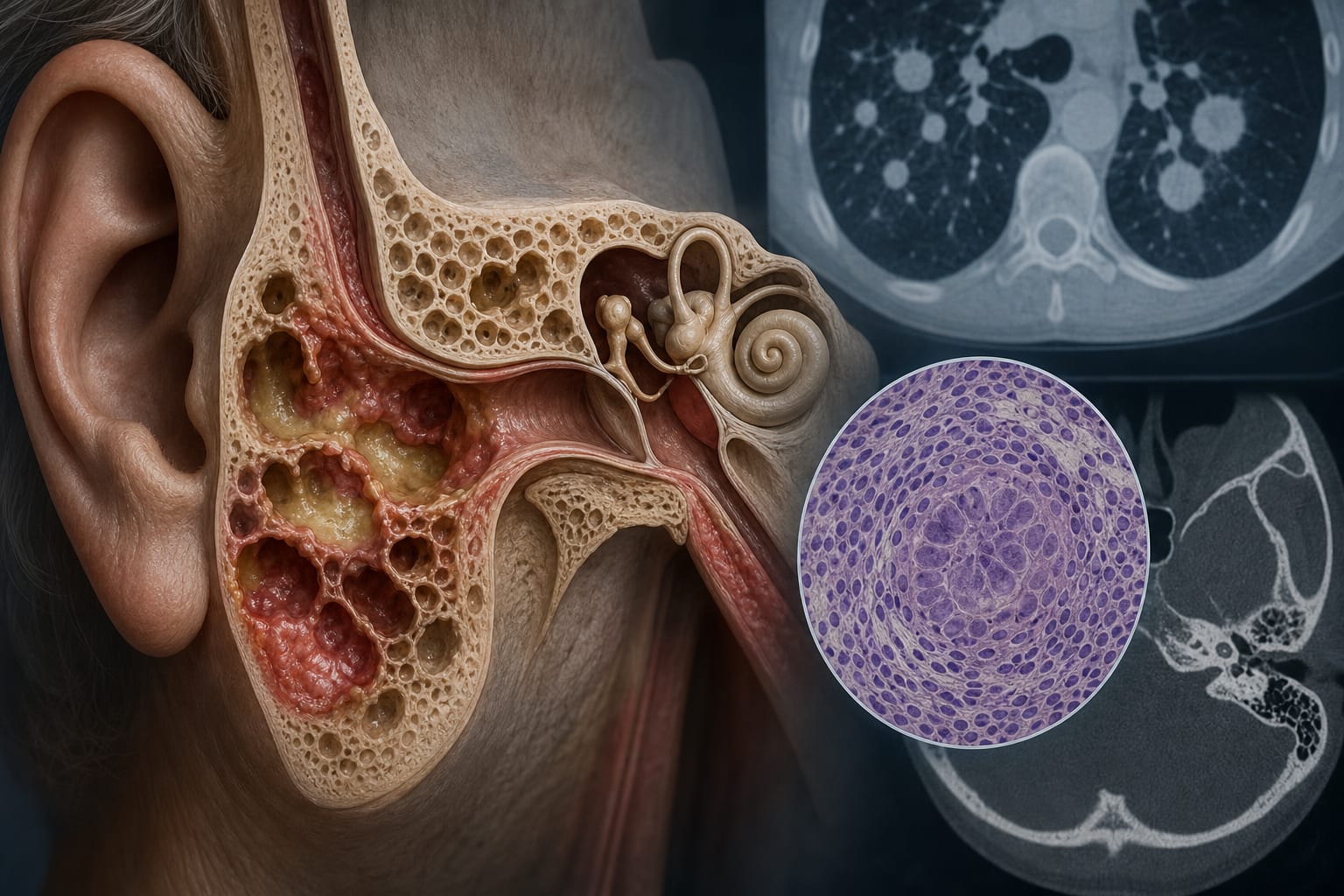
This study investigates the angular trajectory of the vestibular aqueduct (ATVA) in patients with ipsilateral delayed endolymphatic hydrops (DEH) compared to unilateral Ménière’s disease (MD) and control participants.

Background

Delayed endolymphatic hydrops (DEH) and Ménière’s disease (MD) are both characterized by endolymphatic hydrops, but they exhibit distinct clinical features. Understanding the pathological characteristics of the endolymphatic sac (ES) in these conditions is crucial for accurate diagnosis and management. The angular trajectory of the vestibular aqueduct (ATVA) serves as a radiological indicator to differentiate between pathological types of ES in these patients.

Data Highlights

ComparisonFindingp-value
ATVA ≤ 120° in DEH vs MDMore frequent0.002
ATVA ≥ 140° in DEH vs MDLess frequent0.009
Distribution of ATVA in DEH vs ControlNo significant difference0.510
Prevalence of ATVA categories in affected vs non-affected ears in DEHComparable0.568

Key Findings

  • ATVA ≤ 120° was more frequent in DEH-affected ears compared to MD-affected ears.
  • ATVA ≥ 140° was less frequent in DEH-affected ears compared to MD-affected ears.
  • No significant difference in ATVA distribution was found between affected ears of DEH patients and control ears.
  • The prevalence of ATVA categories in affected ears of DEH patients was comparable to that in non-affected ears.
  • Degenerative ES may be the dominant pathological type in ipsilateral DEH patients.

Clinical Implications

The findings indicate the importance of the angular trajectory of the vestibular aqueduct in evaluating patients with ipsilateral DEH.

Conclusion

The study highlights the importance of ATVA as a radiological indicator in distinguishing pathological types of the endolymphatic sac in patients with ipsilateral DEH.

Related Resources & Content

  1. Acta Neuropathologica, 2018 -- Disorders of the Inner Ear Disrupt Sodium-Dependent Ion Transport in Meniere’s Disease
  2. Frontiers in Neurology, 2026 -- Endolymphatic hydrops in patients with migraine: a correlative study
  3. European Radiology, 2024 -- Evaluating Key MRI Features for the Diagnosis of Ménière’s Disease and the Importance of Vestibular Aqueduct Analysis
  4. ICVD Consensus Documents - The Barany Society, 2023
  5. Frontiers, 2023 -- Visualization and clinical relevance of the endolymphatic duct and sac in Ménière's disease
  6. Acta Neuropathologica — Antibody Profiles in Patients with Histologically Characterized Patterns of Multiple Sclerosis
  7. ICVD Consensus Documents - The Barany Society
  8. Visualization and clinical relevance of the endolymphatic duct and sac in Ménière's disease
  9. Frontiers | Degeneration Rather than Hypoplasia May Be the Pathologic Feature of the Endolymphatic Sac in Patients with Ipsilateral Delayed Endolymphatic Hydrops
  10. Abstract Export
  11. Comparative efficacy of intratympanic gentamicin and intratympanic corticosteroid in the treatment of Meniere’s disease: a systematic review and meta-analysis
  12. Comparing Intratympanic to Surgical Management in Refractory Meniere Disease: A Systematic Review Plus Network Meta-analysis - PubMed

Original Source(s)

Related Content