Rethinking the shoe: is CT perfusion the optimal screening tool for acute stroke patients?
-
By
-
Koenraad H. Nieboer
-
October 18, 2023
-
0 min
Clinical Report: Evaluating CT Perfusion as a Screening Tool in Acute Stroke
Overview
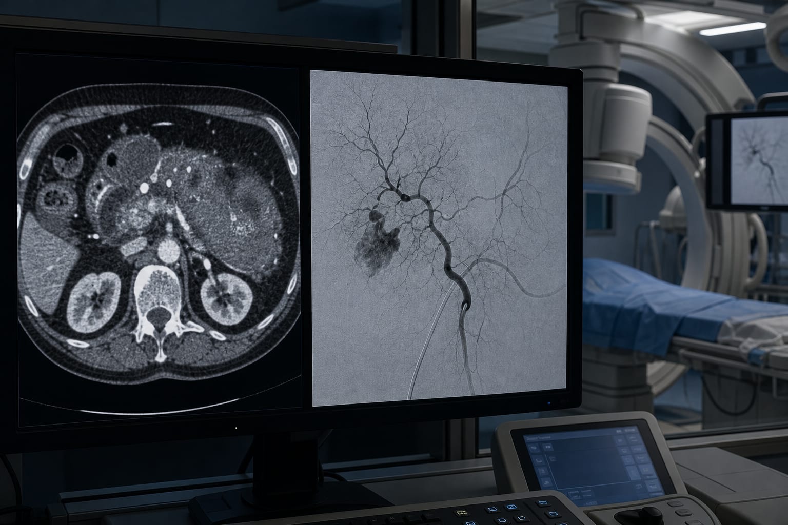
CT perfusion (CTP) imaging has become widely used in acute stroke evaluation due to its availability, despite concerns about radiation exposure and interpretation challenges. While MRI perfusion remains the most accurate and safest method, CTP is frequently employed to identify ischemic core and penumbra, especially for thrombectomy eligibility. Emerging AI tools and multiphase CT angiography may offer alternatives with lower radiation burden.
Background
Brain perfusion imaging is critical in acute stroke management to estimate ischemic core and penumbra volumes, guiding treatment decisions such as intravenous thrombolysis and mechanical thrombectomy. MRI perfusion (MRP) is considered the gold standard due to its accuracy and lack of radiation, but limited availability leads to widespread use of CT perfusion (CTP). European Stroke Organization guidelines recommend perfusion imaging primarily beyond 4.5 hours after symptom onset, yet CTP is often used more broadly to detect candidates for intervention. Automated post-processing tools facilitate CTP interpretation but have limitations including artifacts and lack of standardization.
Data Highlights
Recent studies indicate that patients with large ischemic cores (≥50 mL) or low ASPECT scores (3–5) benefit from endovascular thrombectomy compared to standard care. Distal medium vessel occlusions, often difficult to detect on CTA or MRA, can be better identified with perfusion imaging. AI models applied to non-contrast CT have demonstrated high accuracy in early stroke detection and volume estimation, rivaling human experts and approaching MRI diffusion-weighted imaging accuracy. Multiphase CT angiography can provide temporal and perfusion data with potentially lower radiation exposure than CTP.
Key Findings
- CTP is widely used for acute stroke evaluation due to higher availability compared to MRI perfusion.
- CTP involves higher radiation doses and requires expertise for reliable interpretation; automated software can assist but has limitations.
- There is no standardized thresholding across vendors for ischemic core and penumbra volumes derived from CTP.
- Recent evidence supports thrombectomy in patients with large ischemic cores and distal vessel occlusions, where perfusion imaging aids detection.
- AI applied to non-contrast CT and multiphase CT angiography shows promise as lower-radiation alternatives for stroke detection and perfusion assessment.
- Radiologists must balance the benefits of CTP screening with radiation risks and interpretation challenges, advocating for optimized imaging protocols.
Clinical Implications
Clinicians should be cautious in using CTP as a universal screening tool for acute stroke due to radiation exposure and interpretative complexities. Incorporating AI-enhanced non-contrast CT and multiphase CT angiography may reduce radiation burden while maintaining diagnostic accuracy. Radiologists must remain vigilant about image quality factors and standardization issues when interpreting perfusion studies to guide treatment decisions effectively.
Conclusion
While CTP remains the most accessible perfusion imaging modality in acute stroke, its limitations necessitate reconsideration of its role as a screening tool. Advances in AI and alternative imaging techniques hold promise to improve stroke detection with less radiation, underscoring the need for further research and optimized clinical protocols.
References
- European Stroke Organization Guidelines 2021 -- Management of Acute Ischemic Stroke
- Recent Multicenter Trials 2023 -- Thrombectomy Outcomes in Large Core Infarcts
- Perfusion Imaging Standardization Review 2022 -- Variability in CTP Thresholding
- AI in Stroke Imaging 2023 -- Non-Contrast CT for Early Stroke Detection
- Multiphase CT Angiography Research 2023 -- Perfusion Data from Temporal Phases
- Portable MRI Developments 2024 -- Expanding MRI Access in Stroke Care
This content is an AI-generated, fully rewritten summary based on a published scholarly article. It does not reproduce the original text and is not a substitute for the original publication. Readers are encouraged to consult the source for full context, data, and methodology.