Clinical Report: Investigating Remote Diffusion-Weighted Imaging Lesions in ICH
Overview
This review systematically summarizes the factors influencing remote diffusion-weighted imaging (R-DWI) lesions following spontaneous intracerebral hemorrhage (ICH) and explores their prognostic associations.
Background
Spontaneous intracerebral hemorrhage (ICH) is a leading cause of stroke with high rates of morbidity and mortality. While research has traditionally focused on local lesions, emerging studies highlight the significance of remote ischemic injuries detectable via diffusion-weighted imaging (DWI).
Data Highlights
Study
Frequency of R-DWI Lesions
Study 1
11%
Study 2
50%
Key Findings
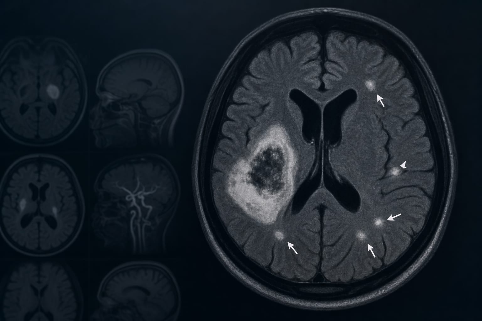
R-DWI lesions are defined as hyperintense areas on DWI located at least 10 mm from the hematoma.
Prevalence of R-DWI lesions in ICH patients ranges from 11% to 50% across studies.
Three main mechanisms for R-DWI lesions include vessel rupture and occlusion, aggressive blood pressure lowering, and inflammatory processes.
Patients with R-DWI lesions may have underlying conditions such as hypertensive arteriopathy or cerebral amyloid angiopathy.
Clinical Implications
Recognizing the presence of R-DWI lesions in ICH patients may guide clinical decision-making and management strategies.
Conclusion
The review emphasizes the importance of understanding R-DWI lesions in ICH.